A collaborative research effort led by Daan van der Vliet and scientists from the Netherlands Institute for Neuroscience, Leiden University, and Utrecht University has identified a critical biological marker that may explain the aggressive trajectory of Multiple Sclerosis (MS) in certain patients. By examining post-mortem brain tissue, the team discovered that an abundance of specialized immune cells, characterized by an accumulation of lipid droplets and referred to as "foamy microglia," is strongly correlated with rapid disease progression and a failure of the brain’s natural repair mechanisms. The study, which utilizes advanced multi-omic analysis, offers a potential breakthrough in the development of predictive biomarkers and personalized therapeutic interventions for one of the most unpredictable neurological conditions.
The Biological Foundation of Multiple Sclerosis
Multiple Sclerosis is a chronic autoimmune disease of the central nervous system (CNS) that affects approximately 2.8 million people worldwide. The pathology of the disease is centered on the destruction of myelin, a fatty substance that forms an insulating sheath around nerve fibers (axons). This insulation is vital for the rapid transmission of electrical impulses between the brain and the rest of the body. When the immune system mistakenly attacks this myelin, the resulting inflammation leads to "lesions" or scarring, which disrupts neural communication.
The clinical manifestation of MS is notoriously heterogeneous. For some patients, the disease follows a relapsing-remitting course (RRMS), where periods of neurological dysfunction are followed by partial or complete recovery. However, for others, the disease transitions into a progressive phase (SPMS) or begins as primary progressive MS (PPMS), leading to a steady decline in motor, sensory, and cognitive functions. Until now, the biological drivers that dictate why some patients experience rapid, debilitating progression while others remain stable for decades have remained largely elusive.
The Role of Microglia: From Protectors to Pathological Drivers
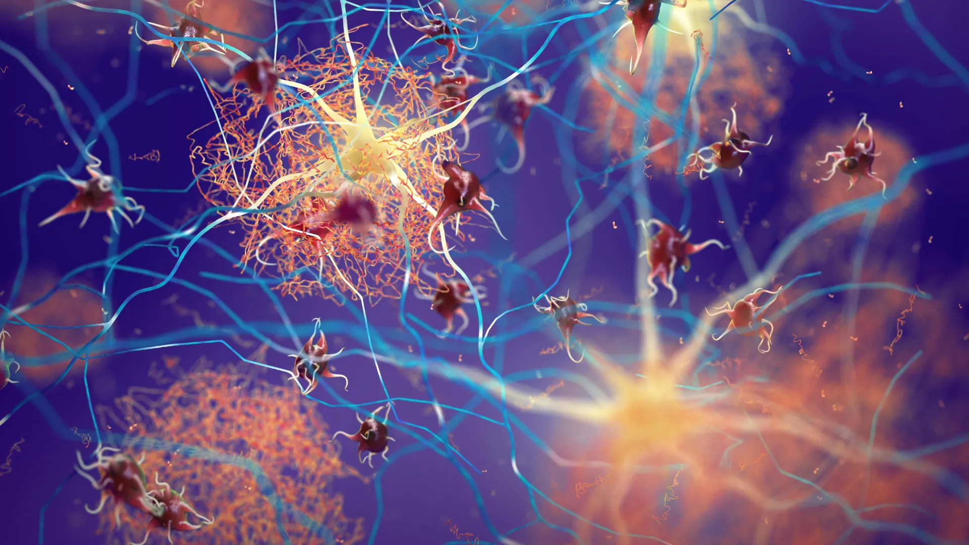
The focus of the new research is the microglia, the resident immune cells of the brain and spinal cord. In a healthy CNS, microglia serve as the primary defense mechanism, constantly surveying the environment for pathogens and cellular debris. When myelin is damaged, microglia are tasked with "cleaning up" the fatty remnants—a process known as phagocytosis—which is a prerequisite for remyelination, the process by which the brain attempts to repair the damaged insulation.
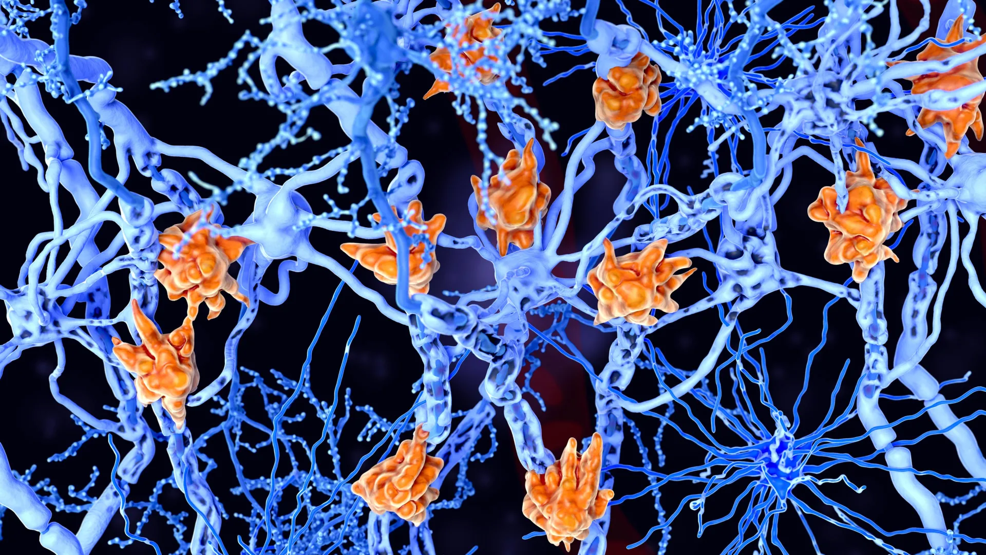
However, the study led by Van der Vliet reveals that in more severe cases of MS, this cleanup process becomes dysfunctional. The researchers observed that in patients with aggressive disease courses, the microglia become overloaded with the lipids they have ingested. This lipid congestion gives the cells a distinctive, bloated appearance under the microscope, leading to the designation "foamy microglia."
"We found that patients with large numbers of these foamy microglia had a more severe disease course more frequently," stated Van der Vliet. The presence of these cells suggests a tipping point where the immune system’s attempt to heal the brain actually contributes to further damage.
The Mechanism of Metabolic Overload
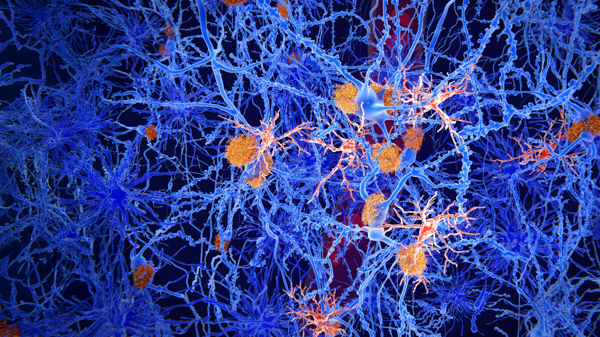
The researchers propose a model of "metabolic exhaustion" to explain the transition of microglia from a regenerative state to a pathological one. Initially, the ingestion of damaged myelin is a beneficial act intended to clear the way for new myelin-producing cells (oligodendrocytes). However, when the volume of damaged myelin exceeds the metabolic capacity of the microglia to process and export the fats, the cells become "clogged."
Once these cells reach a state of lipid overload, their functional profile shifts. Instead of promoting tissue repair, they remain in a chronic inflammatory state. The study indicates that these foamy cells are associated with lesions that show little to no evidence of recovery. "These cells are probably trying to do something good: clearing up damage," Van der Vliet explained. "But they become overloaded, so to speak. As a result, they can no longer effectively contribute to repair."
This failure of the repair cycle creates a self-perpetuating loop of inflammation. The lipid-laden cells release pro-inflammatory signaling molecules that further damage surrounding tissue, leading to more myelin breakdown and, consequently, more lipid debris for the microglia to consume.
Advanced Methodology: Mapping the Molecular Landscape
The findings were made possible through the analysis of brain tissue from 28 deceased MS patients who had donated their organs to the Netherlands Brain Bank (NBB). The NBB is globally recognized for its rigorous protocols in collecting and classifying human brain tissue, providing researchers with a high-resolution window into the lived experience of neurological disease.
To gain a comprehensive understanding of the lesions, the research team employed a "spatial multi-omics" approach. This involved the simultaneous analysis of:
- Transcriptomics: Mapping gene activity within individual cells to see which biological pathways were activated.
- Proteomics: Identifying the proteins being produced by the microglia and surrounding cells.
- Lipidomics: Measuring the specific types of fats present within the foamy cells and the lesion environment.
This detailed mapping allowed the scientists to identify specific molecular signatures that distinguish "active" lesions containing foamy microglia from "silent" or recovering lesions. They found that areas containing foamy cells were enriched with specific lipid species linked to long-term, chronic inflammation.
Van der Vliet emphasized that the synergy between cutting-edge technology and classical pathology was essential. "The technologies are fantastic, but they tell you relatively little if you cannot connect them to pathology in brain tissue. Precisely because brain tissue has been carefully studied and classified for years by the Netherlands Brain Bank, we were able to recognize these abnormal patterns."
Chronology of the Research and Collaborative Framework
The study represents the culmination of several years of interdisciplinary work supported by the Dutch government’s Gravitation programs. These include the Institute for Chemical Immunology (ICI) and the Institute for Chemical NeuroScience (iCNS), which aim to bridge the gap between fundamental chemistry and clinical neurology.
The timeline of the research began with the observation of lipid-rich cells in MS tissue, followed by the development of the multi-omic pipeline necessary to analyze them. By 2022, the team had identified the correlation between foamy microglia and disease severity in a pilot group of patients. The current expanded study confirms these findings across a larger cohort and identifies the specific lipid markers that could eventually be used in clinical settings.
The research also aligns with a growing interest in the pharmaceutical sector regarding the metabolic aspects of MS. Currently, several experimental treatments aimed at modifying lipid metabolism in the brain are being evaluated in clinical studies conducted in collaboration with the pharmaceutical giant Roche. These trials seek to determine if drugs can "jumpstart" the metabolism of microglia, helping them process lipids more efficiently and resume their protective functions.
Toward Personalized Medicine and Future Biomarkers
One of the most significant implications of this discovery is the potential for personalized MS treatment. Currently, doctors often rely on Magnetic Resonance Imaging (MRI) and the presence of "oligoclonal bands" in cerebrospinal fluid (CSF) to diagnose MS and monitor its progression. However, these tools are often reactive rather than predictive.
The discovery of specific fats associated with foamy microglia suggests a new diagnostic pathway. The researchers found evidence that these lipid markers may leak into the cerebrospinal fluid. If future studies can validate a CSF test for these specific lipids, doctors could identify patients at risk of rapid decline much earlier in the disease process.
"That opens the possibility of developing biomarkers in the future that could help doctors identify earlier which patients are at risk of rapid decline—and which treatment would suit them best," Van der Vliet noted. For example, a patient showing high levels of "foamy cell lipids" might be prescribed more aggressive disease-modifying therapies (DMTs) from the outset, rather than following the traditional "step-up" approach where stronger medications are only used after a patient fails on milder ones.
Analysis of Broader Impacts on MS Research
This study contributes to a paradigm shift in how MS is understood. For decades, MS was viewed primarily as a T-cell-mediated autoimmune disease, where the focus was on the infiltration of peripheral immune cells into the brain. While this is still a core component of the disease, the focus is increasingly shifting toward the "innate" immune system of the brain—specifically the microglia.
By identifying the "exhaustion" of microglia as a driver of progression, the research highlights that inflammation and neurodegeneration are not separate processes but are linked by cellular metabolism. This opens the door for a new class of "neuro-reparative" therapies that do not just suppress the immune system but actively support the brain’s internal maintenance systems.
Furthermore, the work underscores the invaluable role of brain donation in medical science. Without the detailed clinical histories and high-quality tissue provided by the donors to the Netherlands Brain Bank, the subtle molecular differences between various types of MS lesions would remain hidden.
Conclusion
The identification of foamy microglia as a hallmark of severe Multiple Sclerosis provides a new lens through which to view the complexity of the disease. By proving that the very cells meant to repair the brain can become agents of its decline when overwhelmed by metabolic waste, Van der Vliet and his colleagues have provided a clear target for future research. As clinical trials with partners like Roche continue, the hope is that the next generation of MS treatments will focus not just on stopping damage, but on clearing the way for the brain to heal itself. For millions of patients living with the uncertainty of MS, these findings offer a roadmap toward a future where disease progression is no longer a mystery, but a manageable and predictable aspect of their care.