The discovery of a singular molecular mechanism that governs the transition of blood stem cells from a dormant state to an active, regenerative powerhouse marks a significant milestone in the field of regenerative medicine. In a comprehensive preclinical study led by investigators at Weill Cornell Medicine, researchers have identified a specific DNA transcription-regulating protein, FLI-1, as the essential "switch" required for hematopoietic stem cells to begin the process of rapid multiplication and tissue replacement. This finding, published in the journal Nature Immunology, offers a potential paradigm shift in how clinicians approach bone marrow transplants and gene therapies, particularly for patients with limited donor options or compromised cellular health.
Unlocking the Regenerative Potential of Hematopoietic Stem Cells
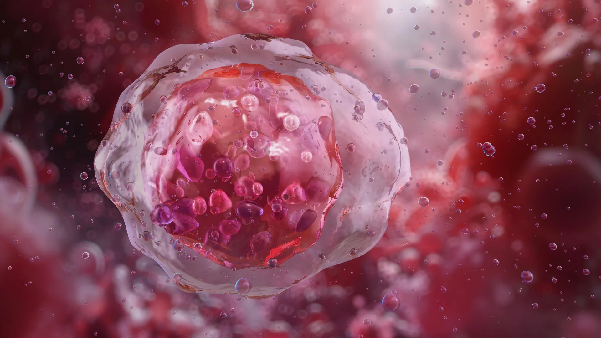
Stem cells serve as the fundamental biological building blocks for nearly all human tissues. Within the blood-forming system, these hematopoietic stem cells (HSCs) are primarily found within the protective environment of the bone marrow. Under normal physiological conditions, these cells exist in a state of "quiescence"—a deep metabolic slumber characterized by slow division and minimal activity. This dormant state is a protective evolutionary mechanism, designed to preserve the long-term viability of the stem cell pool by shielding it from genetic mutations and metabolic stress.
However, when the body experiences significant injury, blood loss, or the depletion of immune cells, these quiescent cells must "wake up." This transition to an activated state allows them to migrate into the bloodstream, multiply at an exponential rate, and eventually differentiate into mature, functional red blood cells, white blood cells, and platelets. The Weill Cornell study has successfully mapped the genetic pathways that facilitate this transition, focusing on the role of FLI-1 in orchestrating the complex interplay between the stem cells and their surrounding environment.
The Critical Role of FLI-1 in Stem Cell Activation
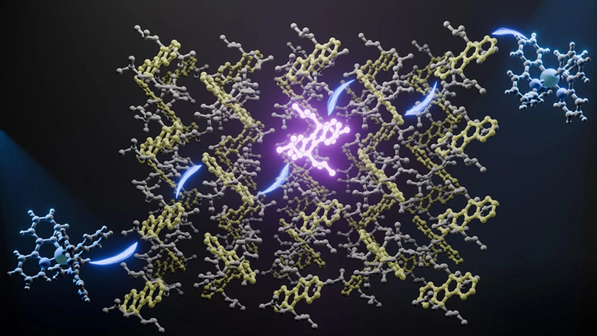
The research team, utilizing advanced single-cell profiling and computational analysis, sought to understand the stark differences in gene expression between dormant and active blood stem cells. Through their investigation, they pinpointed FLI-1, a transcription factor protein known to regulate the activity of thousands of downstream genes. The study demonstrated that FLI-1 acts as the primary driver of the regenerative process. In its absence, blood stem cells remain trapped in a state of hibernation, unable to respond to the body’s demands for new tissue.
One of the most significant aspects of the discovery is how FLI-1 facilitates communication between the stem cells and the "vascular niche." This niche consists of specialized endothelial cells that line the blood vessels within the bone marrow. The researchers found that FLI-1 restores the stem cells’ ability to connect and co-adapt with these endothelial cells. This interaction is not merely a passive proximity; it is a dynamic signaling exchange that provides the stem cells with the necessary cues to begin expansion. By transiently producing FLI-1 in adult bone marrow stem cells, the researchers were able to "prime" these cells, significantly improving their ability to engraft and restore the blood supply when transplanted into a new host.
Addressing the Safety Concerns of Genetic Overexpression
While the potency of FLI-1 as a regenerative agent is clear, its power comes with inherent risks. In clinical oncology, mutations that lead to the chronic overactivity of FLI-1 are well-documented drivers of certain types of leukemia. The challenge for the research team was to harness the regenerative benefits of the protein without triggering malignant transformations.
To solve this, the investigators developed an innovative delivery method inspired by the technology used in modified mRNA-based vaccines. Instead of permanently altering the genetic makeup of the stem cells, the team introduced FLI-1 transiently. By using modified mRNA, they were able to stimulate the production of the protein for only a few days. This short window of activity was sufficient to "wake up" the cells and initiate the regenerative cycle without providing the sustained signaling that could lead to cancer.
Dr. Tomer Itkin, co-first author of the study and current director of Tel Aviv University’s Neufeld Cardiovascular Research Institute, emphasized the success of this approach. According to Dr. Itkin, the stem cells primed with FLI-1 modified mRNA effectively exited hibernation, expanded, and achieved durable engraftment in recipient hosts without any evidence of oncogenic activity. This transient activation model provides a safe and scalable framework for clinical application.
The Evolution of Bone Marrow Transplantation and Gene Therapy
The implications of this research are particularly profound for bone marrow transplantation, a procedure that has been a cornerstone of treatment for blood cancers and immune disorders for decades. In many cases, the success of a transplant depends on the quantity and quality of the donor’s stem cells. Patients who have undergone intensive chemotherapy or radiation often have "exhausted" stem cell pools that are difficult to activate. By using FLI-1 to prime these cells, doctors could potentially improve the efficiency of transplants even when the donor supply is limited.
Furthermore, the discovery holds great promise for the burgeoning field of gene therapy. For conditions such as beta-thalassemia or sickle cell anemia, patients’ own stem cells are harvested, genetically corrected in a laboratory setting, and then re-infused. However, the process of expanding these corrected cells outside the body is often slow and inefficient. Integrating a transient FLI-1 activation step could streamline this process, ensuring that the laboratory-grown cells are in an optimal state for engraftment once they return to the patient’s body.
Deciphering the Potency of Umbilical Cord Blood
The study also shed light on a long-standing mystery in hematology: why stem cells derived from umbilical cord blood generally possess a higher regenerative capacity than those harvested from adult bone marrow. The research team found that this disparity is directly linked to the levels of FLI-1 activity. Umbilical cord-derived stem cells naturally exhibit higher levels of FLI-1, which explains their superior ability to interact with the vascular niche and rapidly replenish the blood system. By understanding this natural mechanism, the researchers have essentially found a way to "rejuvenate" adult stem cells, giving them the regenerative vigor typically associated with neonatal cells.
Computational Breakthroughs and the Future of Regenerative Medicine
The success of the study was predicated on extensive computational analysis. Sean Houghton, a co-first author and bioinformatics analyst, noted that the research clarified that stem cell activity is not an autonomous process. Instead, it is a collaborative effort between the stem cells and the signals provided by the endothelial cells in the vascular niche. The study utilized bioinformatics to decipher how FLI-1 integrates with known signaling pathways that drive cell survival and self-renewal.
This systems-biology approach allowed the team to move beyond identifying single genes and instead understand the entire "network" of activation. "We showed that stem cell activity… depends instead on signaling and adaptability between the two," Houghton explained, referring to the stem cells and their environment. This insight reinforces the growing understanding in precision medicine that the microenvironment, or "niche," is just as important as the cells themselves.
Funding and Institutional Collaboration
The research was a collaborative effort involving several leading institutions and was supported by significant federal funding. The National Heart, Lung, and Blood Institute, the National Institute of Diabetes and Digestive and Kidney Diseases, and the National Institute of Allergy and Infectious Diseases—all branches of the National Institutes of Health (NIH)—provided the primary financial support through various grants.
Additional backing was provided by the Hartman Institute for Therapeutic Organ Regeneration, the Ansary Stem Cell Institute, and the Selma and Lawrence Ruben Daedalus Fund for Innovation at Weill Cornell Medicine. Dr. Shahin Rafii, the study’s senior author and a prominent figure in regenerative medicine, noted that the team’s next steps involve scaling up the modified mRNA-based method for further preclinical development, with the ultimate goal of transitioning into human clinical trials.
As the medical community moves toward more personalized and efficient treatments for blood disorders, the identification of FLI-1 as a master regulator of stem cell activation provides a clear roadmap. By mastering the "on-off" switch of the blood’s regenerative system, researchers are paving the way for a new era of stable, safe, and long-term blood production for patients worldwide. This advancement not only enhances our understanding of cellular biology but also offers tangible hope for those facing life-threatening hematologic conditions.