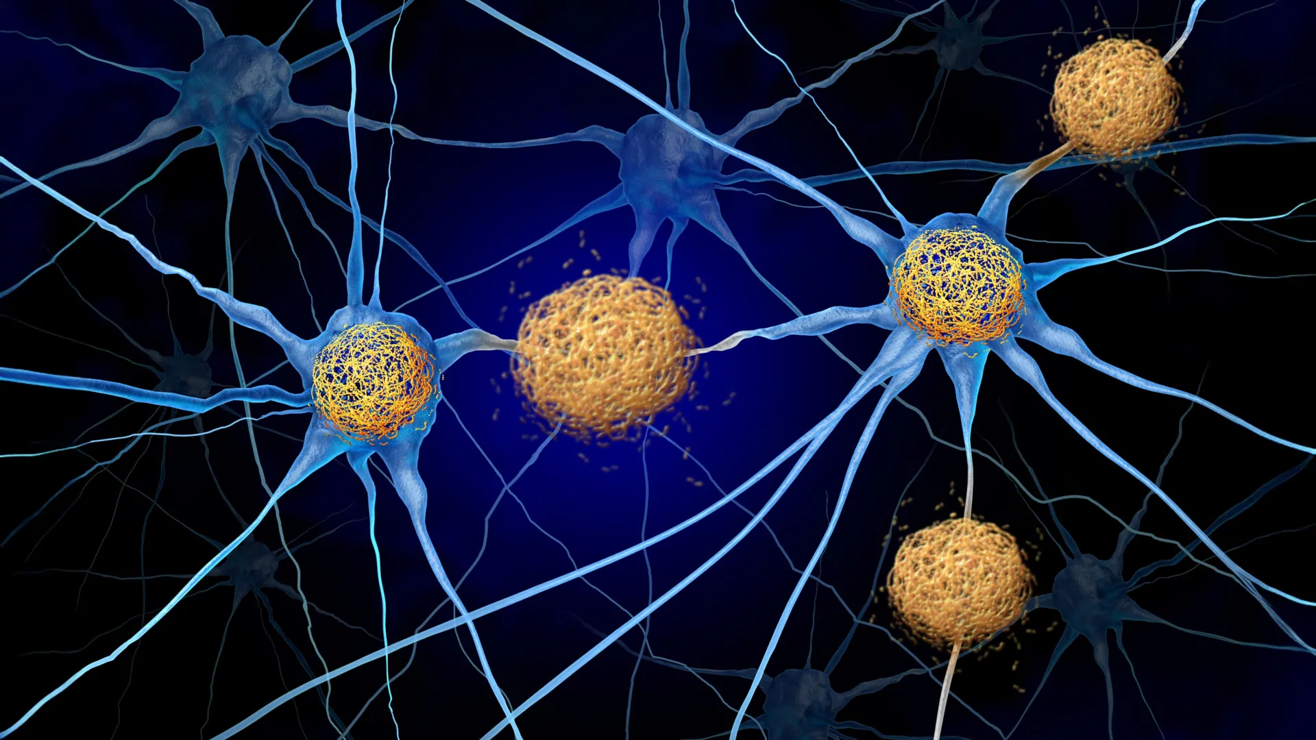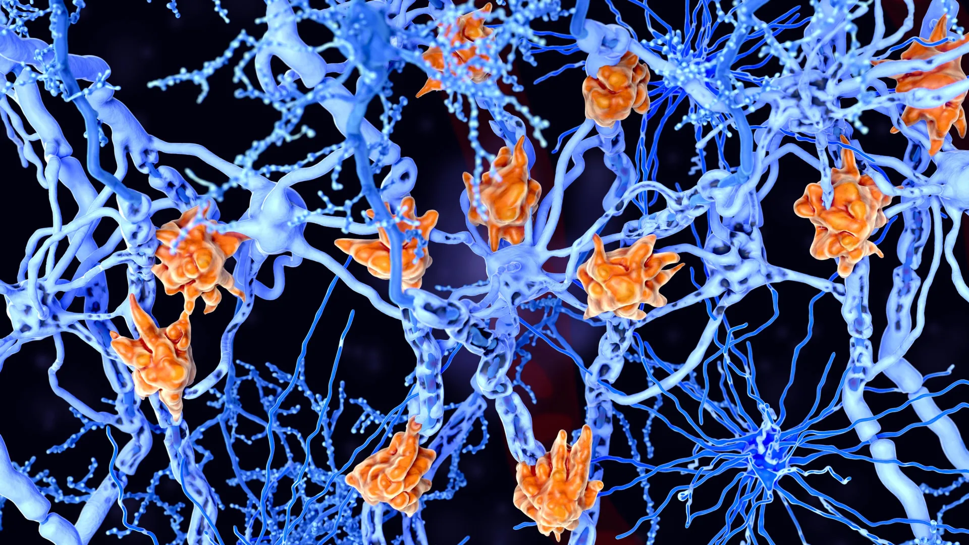
A collaborative research effort led by Daan van der Vliet and a multidisciplinary team from the Netherlands Institute for Neuroscience, Leiden University, and Utrecht University has uncovered a critical biological mechanism that explains the variance in disease severity among those living with multiple sclerosis (MS). By analyzing post-mortem brain tissue, the researchers identified a specific population of immune cells—microglia—that become overloaded with lipid droplets, leading to a state described as "foamy." This cellular transformation appears to be a hallmark of more aggressive forms of the disease, potentially offering a roadmap for new diagnostic biomarkers and targeted therapeutic interventions.
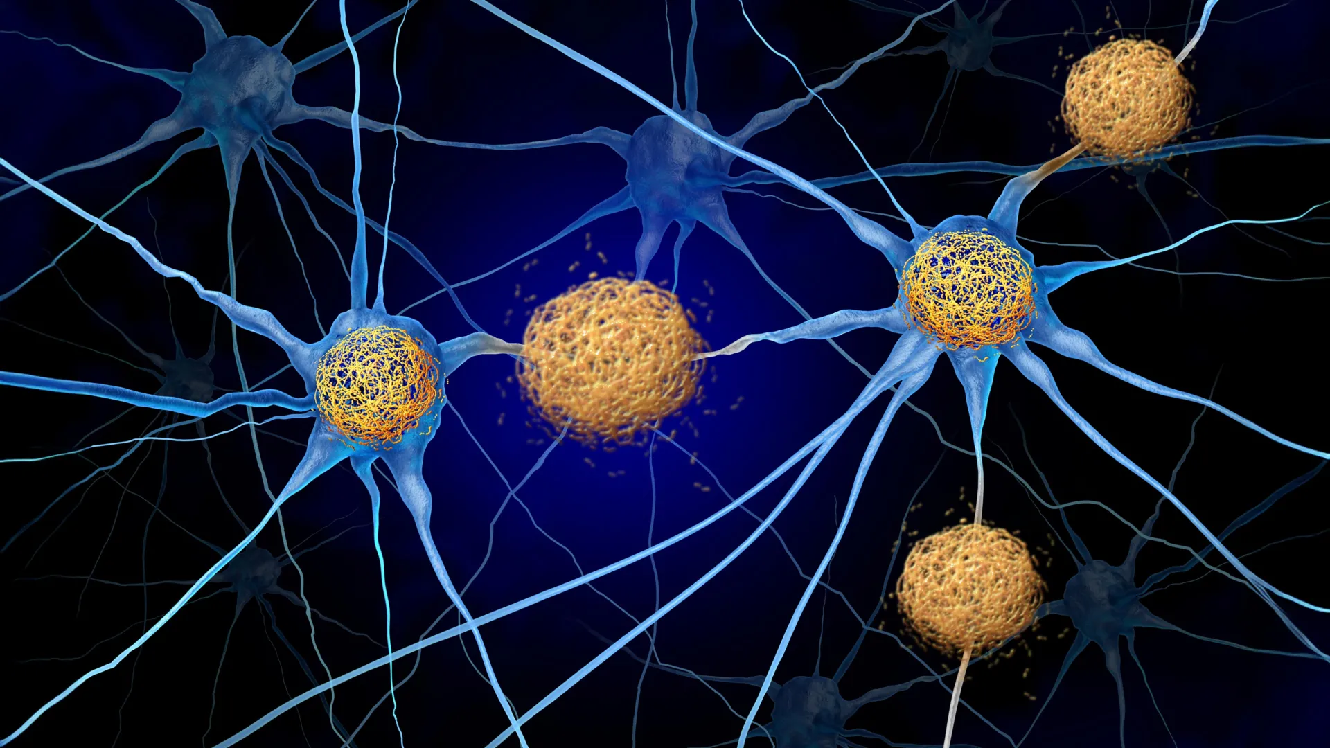
Multiple sclerosis is a chronic autoimmune condition characterized by the immune system’s misguided attack on the central nervous system. Specifically, the body targets myelin, the fatty substance that insulates nerve fibers (axons). When myelin is damaged, the transmission of electrical impulses between the brain and the rest of the body is disrupted, resulting in symptoms ranging from numbness and vision loss to paralysis and cognitive decline. While the fundamental mechanics of MS have been understood for decades, the reason why some patients experience a slow, manageable disease course while others face rapid, debilitating progression has remained one of neurology’s most persistent mysteries.
The Role of Microglia in Neurodegeneration
The study’s focus on microglia represents a shift in how researchers view the progression of MS. Microglia are the resident immune cells of the brain and spinal cord, acting as a specialized cleanup crew. Under normal conditions, they are essential for maintaining neural health, scavenging for pathogens, and removing cellular debris. In the context of MS, their primary task is to clear away the fragments of damaged myelin to make way for remyelination—the process by which the brain attempts to repair its own insulation.
However, the findings published by Van der Vliet and his colleagues suggest that this cleanup process can become a double-edged sword. When the breakdown of myelin is too extensive or occurs too rapidly, the microglia ingest more fat than they can metabolize. These cells then transform into "foamy microglia," named for their distinctive appearance under a microscope when packed with lipid droplets.
"We found that patients with large numbers of these foamy microglia had a more severe disease course more frequently," stated Van der Vliet. The presence of these cells indicates a tipping point where the brain’s natural repair mechanism fails, transitioning from a protective state to one that actively contributes to the disease’s pathology.
A Chronology of Discovery and Methodological Rigor
The path to this discovery was paved by decades of meticulous tissue preservation and the evolution of "multi-omic" technology. The study relied on the analysis of brain tissue from 28 deceased MS patients who had donated their organs to the Netherlands Brain Bank (NBB). Since its inception in 1985, the NBB has become a global cornerstone for neurological research, providing scientists with high-quality, clinically categorized human tissue that allows for a level of detail impossible to achieve through animal models or standard imaging.
The research team utilized a suite of advanced analytical techniques, including spatial transcriptomics and high-resolution lipidomics. This allowed them to map gene activity and protein expression while simultaneously identifying the specific types of fats present within individual MS lesions.
Historically, MS research was limited to looking at "active" versus "inactive" lesions. The timeline of this field shows a progression from simple cellular staining in the mid-20th century to the molecular mapping used today. By 2010, researchers had begun to suspect that the metabolic state of immune cells was as important as their presence. This latest study confirms that hypothesis by demonstrating that the molecular "signature" of a lesion changes drastically once foamy microglia become the dominant cell type.
The Failure of the "Cleanup" Mechanism
When microglia become overloaded with lipids, they enter a state of metabolic exhaustion. The study reveals that these cells are not merely passive observers of damage; their inability to process fat triggers a cascade of negative effects. Instead of promoting tissue repair, foamy microglia begin to secrete pro-inflammatory signaling molecules.
"These cells are probably trying to do something good: clearing up damage," Van der Vliet explained. "But they become overloaded, so to speak. As a result, they can no longer effectively contribute to repair."
This finding challenges the traditional view that inflammation in MS is a primary event driven solely by T-cells and B-cells from the peripheral immune system. Instead, it suggests a more complex, cyclical process: initial myelin damage leads to microglial overload, which in turn causes metabolic failure, leading to chronic inflammation and the prevention of any meaningful recovery. The researchers found that areas of the brain rich in foamy microglia were also enriched with specific long-chain fatty acids and inflammatory lipids, creating a toxic environment for surviving neurons.
Supporting Data and Statistical Significance
The study’s data highlights a clear correlation between microglial lipid content and clinical outcomes. Patients categorized as having "rapidly progressing MS" showed a significantly higher density of foamy microglia in both the white and gray matter compared to those with a "benign" or slow-moving disease course.
Furthermore, the molecular analysis identified specific lipid species—particularly certain ceramides and oxidized phospholipids—that were uniquely present in the lesions of the most severely affected patients. These lipids are known to be potent drivers of inflammation. The researchers noted that the presence of these fats coincided with a decrease in the expression of genes associated with "remyelination" and "trophic support," suggesting that once a lesion becomes "foamy," the window for natural repair effectively closes.
Strategic Implications for Personalized Medicine
The identification of these fat-laden cells offers a new frontier for personalized MS treatment. Currently, most MS therapies are immunomodulators designed to reduce the frequency of relapses by suppressing the immune system. While effective for relapsing-remitting MS (RRMS), these treatments often fail to halt the progression of disability in patients with progressive forms of the disease.
The discovery of foamy microglia points toward two potential breakthroughs:
- Diagnostic Biomarkers: The researchers found evidence that the specific fats associated with foamy microglia may "leak" into the cerebrospinal fluid (CSF). If a diagnostic test can be developed to detect these lipid signatures in living patients, doctors could identify those at risk for rapid decline much earlier than is currently possible. This would allow for more aggressive intervention before irreversible damage occurs.
- Metabolic Targeting: Instead of just suppressing the immune system, future therapies could focus on helping microglia process fats more efficiently. By targeting lipid metabolism pathways, it may be possible to prevent microglia from becoming "overloaded," thereby maintaining their ability to repair the brain.
Official Responses and Collaborative Outlook
The scientific community has responded to the findings with cautious optimism. Independent neurologists have noted that while the study was conducted on post-mortem tissue, the correlation with clinical history is robust. The collaboration with pharmaceutical giant Roche underscores the clinical relevance of the work. Roche is currently involved in evaluating experimental treatments that target chronic MS lesions and lipid metabolism, and the data from the Van der Vliet study provides a biological rationale for these efforts.
The research was also bolstered by the Gravitation programs: the Institute for Chemical Immunology (ICI) and the Institute for Chemical NeuroScience (iCNS). These programs represent a strategic investment by the Dutch government to bridge the gap between chemistry and clinical medicine.
"Today we have incredibly sophisticated techniques that can map the brain in great detail," Van der Vliet noted. "The technologies are fantastic, but they tell you relatively little if you cannot connect them to pathology in brain tissue." This sentiment highlights the ongoing necessity of biobanking and the integration of diverse scientific disciplines—from lipid chemistry to clinical neurology.
Broader Impact on Neurodegenerative Research
The implications of this study extend beyond multiple sclerosis. Lipid accumulation in immune cells is a phenomenon observed in other neurodegenerative conditions, including Alzheimer’s disease and Amyotrophic Lateral Sclerosis (ALS). The "foamy" phenotype seen in MS microglia may be a universal indicator of a failed immune response in the central nervous system.
By shifting the focus from the quantity of inflammation to the quality and metabolic state of the immune cells involved, this research opens a new chapter in neuroimmunology. The goal is no longer just to stop the immune system from attacking the brain, but to ensure that the brain’s own defense and repair systems remain functional under the stress of disease.
As the medical community moves toward a more nuanced understanding of MS, the work of Van der Vliet and his colleagues serves as a reminder that the path to a cure lies in understanding the delicate balance of cellular metabolism. The transition of a microglial cell from a "helper" to a "hindrance" is a subtle biological shift, but for the millions of people living with MS, understanding and reversing that shift could mean the difference between a life of mobility and one of profound disability. Future studies will now focus on validating the CSF biomarkers in larger cohorts of living patients, bringing the promise of personalized MS care closer to clinical reality.