The human skeletal system is a marvel of dynamic structural engineering, continuously remodeling itself in response to the physical demands placed upon it. For generations, medical science has understood the empirical correlation between physical activity and bone density; exercise builds robust, fracture-resistant bones, whereas a sedentary lifestyle leads directly to skeletal deterioration. However, the precise molecular mechanisms by which mechanical forces are converted into biological signals—specifically how bones "know" they are moving—have remained largely elusive.
Now, a team of researchers from the Department of Medicine at the School of Clinical Medicine, LKS Faculty of Medicine, University of Hong Kong (HKUMed), has successfully identified the biological process that bridges physical movement and bone maintenance. By isolating a specific protein that functions as the body’s internal exercise sensor, the research team has unlocked a pathway that could revolutionize the prevention and treatment of osteoporosis. Published in the peer-reviewed journal Signal Transduction and Targeted Therapy, this breakthrough opens the tangible possibility of developing pharmacological interventions that mimic the osteoprotective benefits of physical activity, offering a vital lifeline for millions of individuals who are physically incapable of exercise.
The Global Burden of Osteoporosis and Age-Related Bone Loss
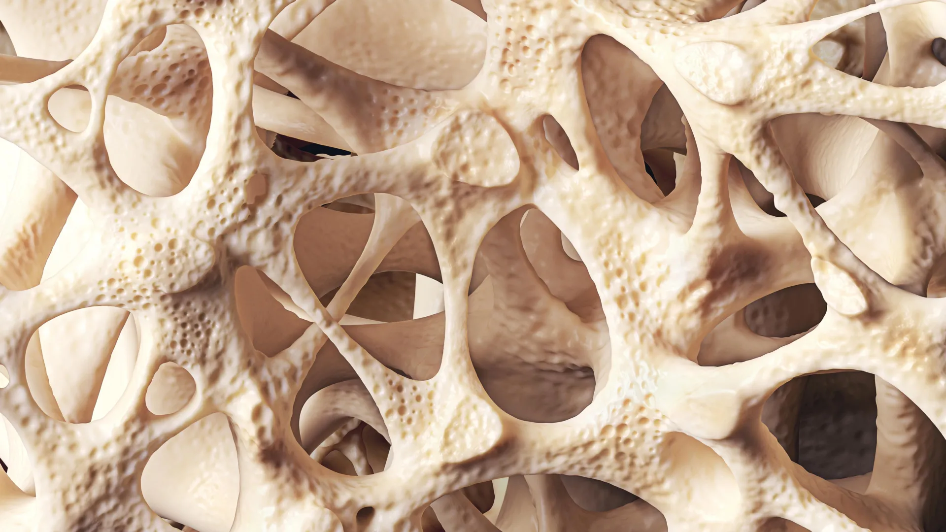
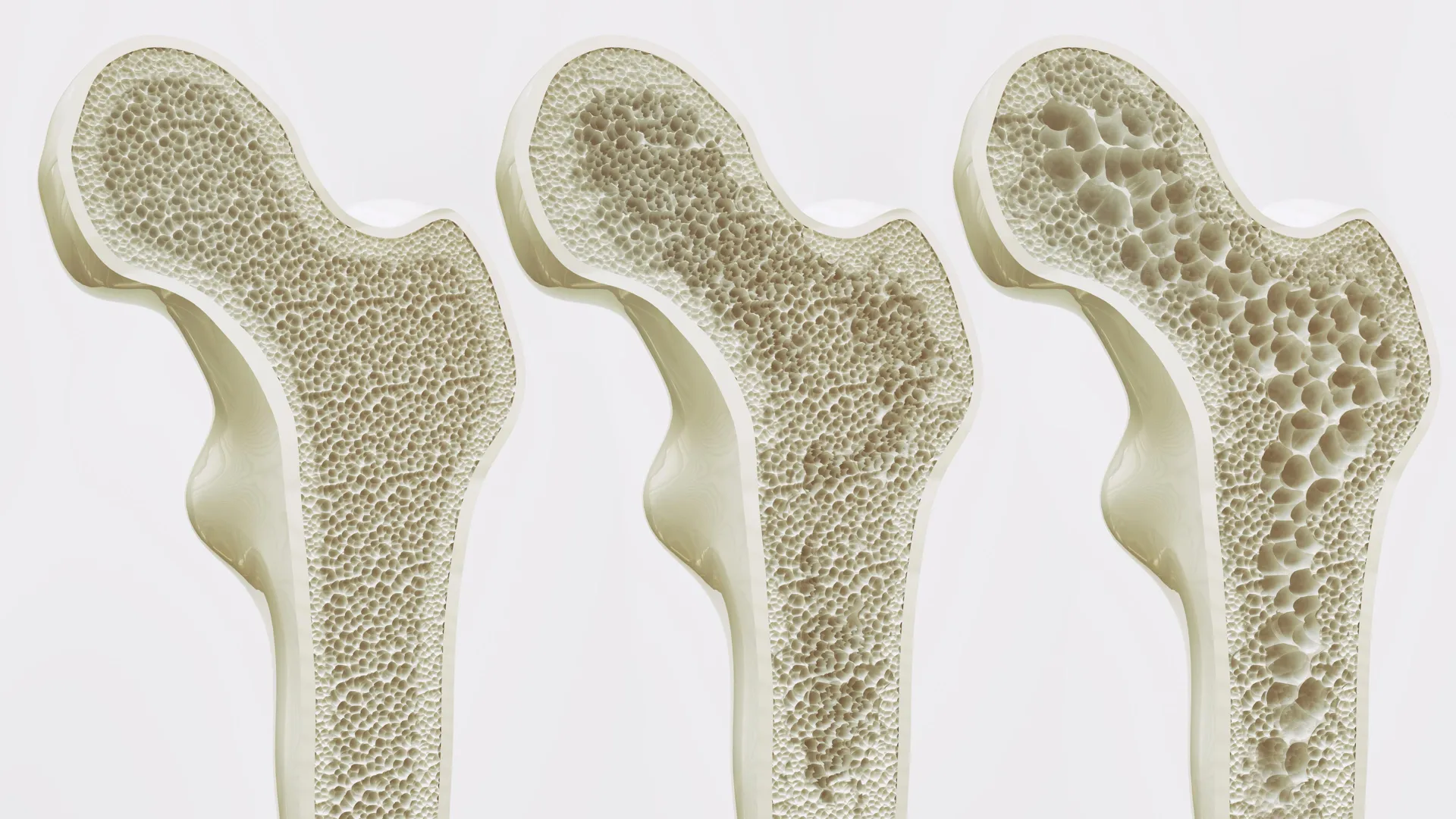
To fully appreciate the significance of the HKUMed discovery, one must examine the staggering global public health crisis posed by osteoporosis. Often described as a silent disease because bone loss occurs without symptoms until a fracture happens, osteoporosis is characterized by systemic skeletal deterioration, low bone mass, and micro-architectural degradation of bone tissue.
According to epidemiological data from the World Health Organization (WHO), approximately one in three women and one in five men over the age of 50 will experience an osteoporotic fracture in their lifetime. These fractures most frequently occur in the hip, spine, and wrist, leading to catastrophic losses in mobility, chronic pain, and a severely diminished quality of life. In many cases, a major osteoporotic fracture—particularly of the hip—initiates a downward health spiral, with significant one-year mortality rates among elderly patients and a permanent loss of independent living.
The socio-economic and demographic pressures of this condition are acute in densely populated urban centers with rapidly aging demographics, such as Hong Kong. Local health statistics indicate that osteoporosis affects an alarming 45% of women and 13% of men aged 65 and older. As global life expectancies continue to rise, healthcare systems face mounting strains from fracture-related hospitalizations, long-term rehabilitation, and specialized nursing care. Current therapeutic options, while helpful, are largely limited to pharmacological agents that either slow bone resorption or modestly stimulate bone formation, yet they frequently fail to address the root cellular imbalance caused by physical inactivity and aging.
The Cellular Tug-of-War Inside the Bone Marrow
To understand why bones grow increasingly fragile with age—and why this deterioration accelerates drastically when a person becomes bedridden or sedentary—researchers examined the microenvironment of the bone marrow.
At the center of skeletal maintenance are mesenchymal stem cells (MSCs) residing within the bone marrow. These multipotent stromal cells possess the remarkable capacity to differentiate into various cell lineages, primarily osteoblasts (bone-forming cells) or adipocytes (fat cells). In a young, healthy individual, mechanical loading from daily movement, gravity, and exercise exerts physical forces on the bone matrix, signaling MSCs to differentiate into osteoblasts, thereby maintaining or increasing bone mineral density.
However, the natural aging process disrupts this delicate cellular equilibrium. Over time, systemic biological shifts alter the mechanotransduction pathways of the bone marrow. Instead of developing into bone-forming osteoblasts, an increasing proportion of mesenchymal stem cells undergo adipogenesis, differentiating into fat cells.
This accumulation of fat within the bone marrow is far more than a passive byproduct of aging; it is an active driver of skeletal decline. As adipose tissue builds up inside the restricted spatial confines of the bone marrow cavity, it physically crowds out healthy bone-forming tissues. Furthermore, marrow adipose tissue secretes paracrine factors that inhibit osteoblastogenesis while stimulating osteoclast activity (cells that break down bone tissue). This creates a vicious, self-reinforcing cycle of structural deterioration that standard anti-osteoporotic drugs struggle to reverse once advanced.
Piezo1: The Molecular Switchboard of Mechanical Force
Seeking to interrupt this pathological cycle, the HKUMed research team—in a collaborative effort involving international institutions—focused on identifying the precise molecular machinery responsible for translating physical forces into biochemical signals within mesenchymal stem cells.
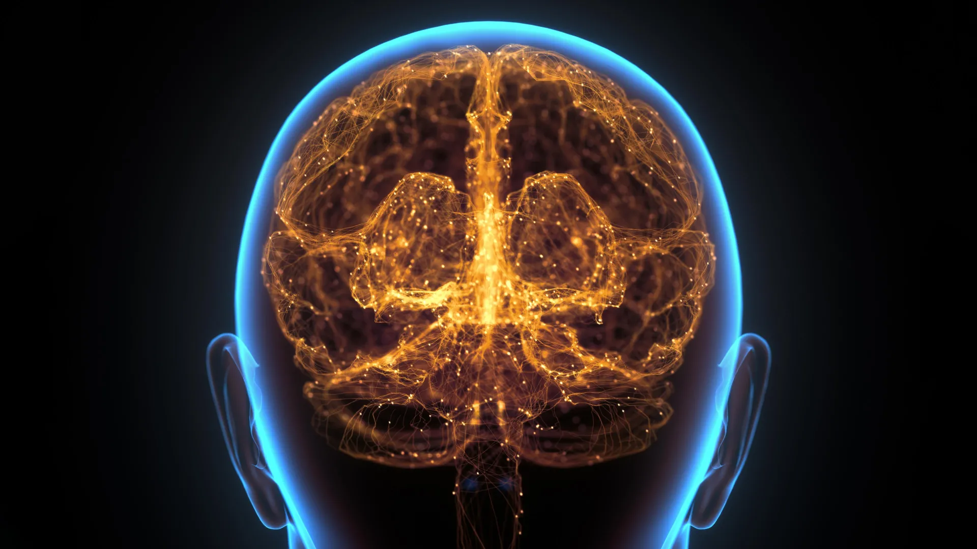
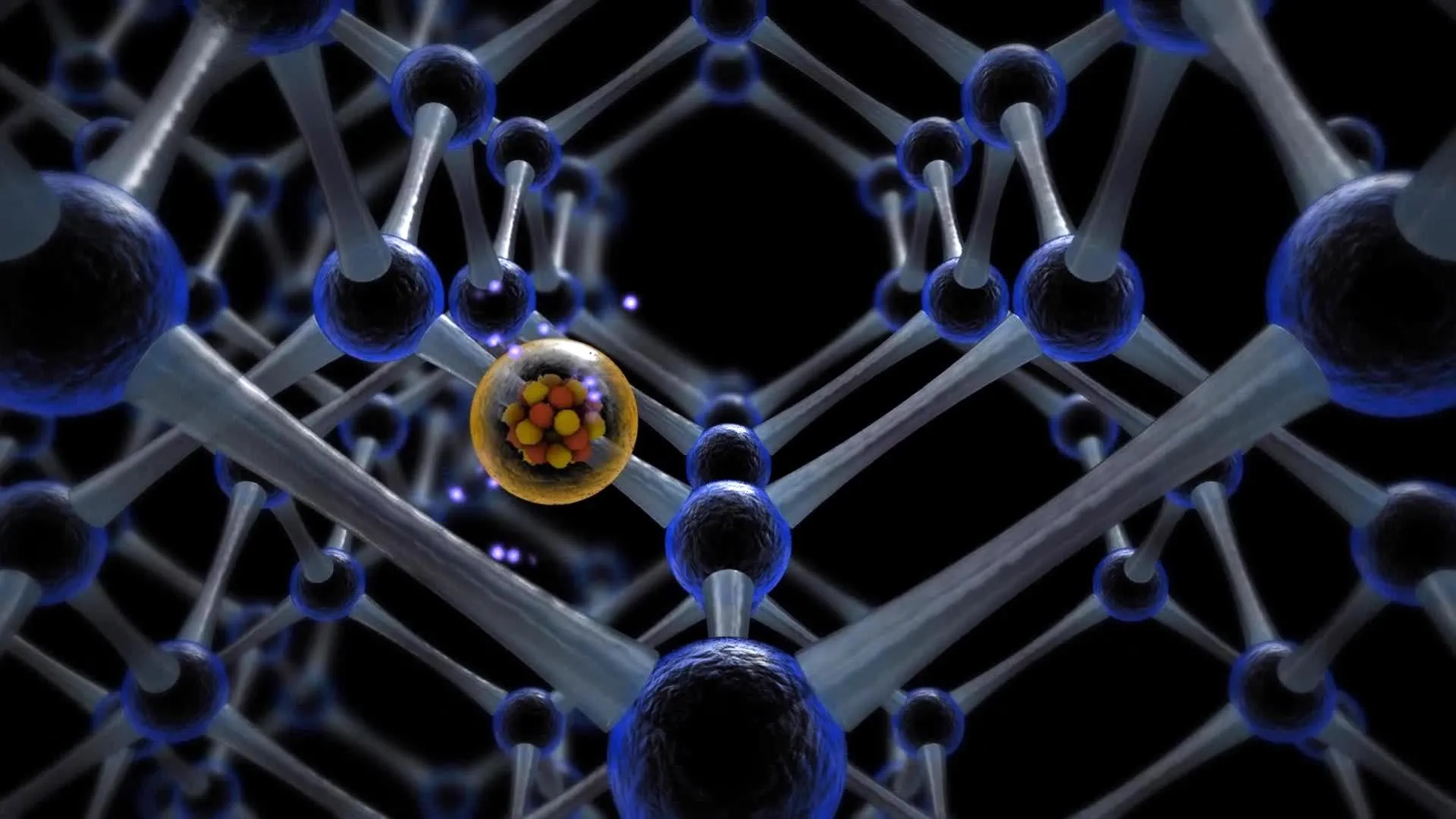
Utilizing a combination of advanced mouse models and human stem cell cultures, the investigators discovered that a specific mechanosensitive ion channel protein, designated as Piezo1, plays a pivotal role. Located on the surface membrane of bone marrow mesenchymal stem cells, Piezo1 acts as a microscopic biological antenna, detecting the physical forces and fluid shear stress generated during movement and exercise.
When physical forces activate Piezo1, the protein triggers a cascade of intracellular signaling events. Experimentally, the researchers observed that activation of Piezo1 in murine models effectively suppressed adipogenesis, steering mesenchymal stem cells away from fat production and routing them toward osteoblast differentiation, thereby promoting new bone formation.
Conversely, when the Piezo1 protein was genetically deleted or impaired, the cellular response inverted entirely. Stem cells preferentially differentiated into fat cells, accelerating bone mass loss. Furthermore, the absence of functional Piezo1 triggered the pathological secretion of specific inflammatory signaling molecules—namely Ccl2 and lipocalin-2. These inflammatory cues further exacerbated fat accumulation within the bone marrow microenvironment while actively suppressing bone growth. Crucially, the research team demonstrated that pharmacologically or genetically blocking these inflammatory signals could help restore healthier bone conditions, highlighting multiple potential points for therapeutic intervention.
Translating Science: The Promise of Exercise Mimetics
The identification of Piezo1 and its downstream signaling pathways represents a paradigm shift in how biomedical researchers conceptualize the prevention and treatment of bone loss. By decoding the molecular translation of movement into skeletal strength, scientists have moved past the empirical observation that exercise is good for bones and identified the exact biological buttons that physical activity pushes.
"We have essentially decoded how the body converts movement into stronger bones," stated Professor Xu Aimin, Director of the State Key Laboratory of Pharmaceutical Biotechnology, Chair Professor in the Department of Medicine, and Rosie T T Young Professor in Endocrinology and Metabolism at HKUMed, who led the investigation. "We have identified the molecular exercise sensor, Piezo1, and the signaling pathways it controls. This gives us a clear target for intervention. By activating the Piezo1 pathway, we can mimic the benefits of exercise, effectively tricking the body into thinking it is exercising, even in the absence of movement."
This breakthrough carries profound implications for clinical populations for whom physical therapy or voluntary exercise is an impossibility. Dr. Wang Baile, Research Assistant Professor in the Department of Medicine at HKUMed and co-leader of the study, emphasized the translational potential for vulnerable demographics.
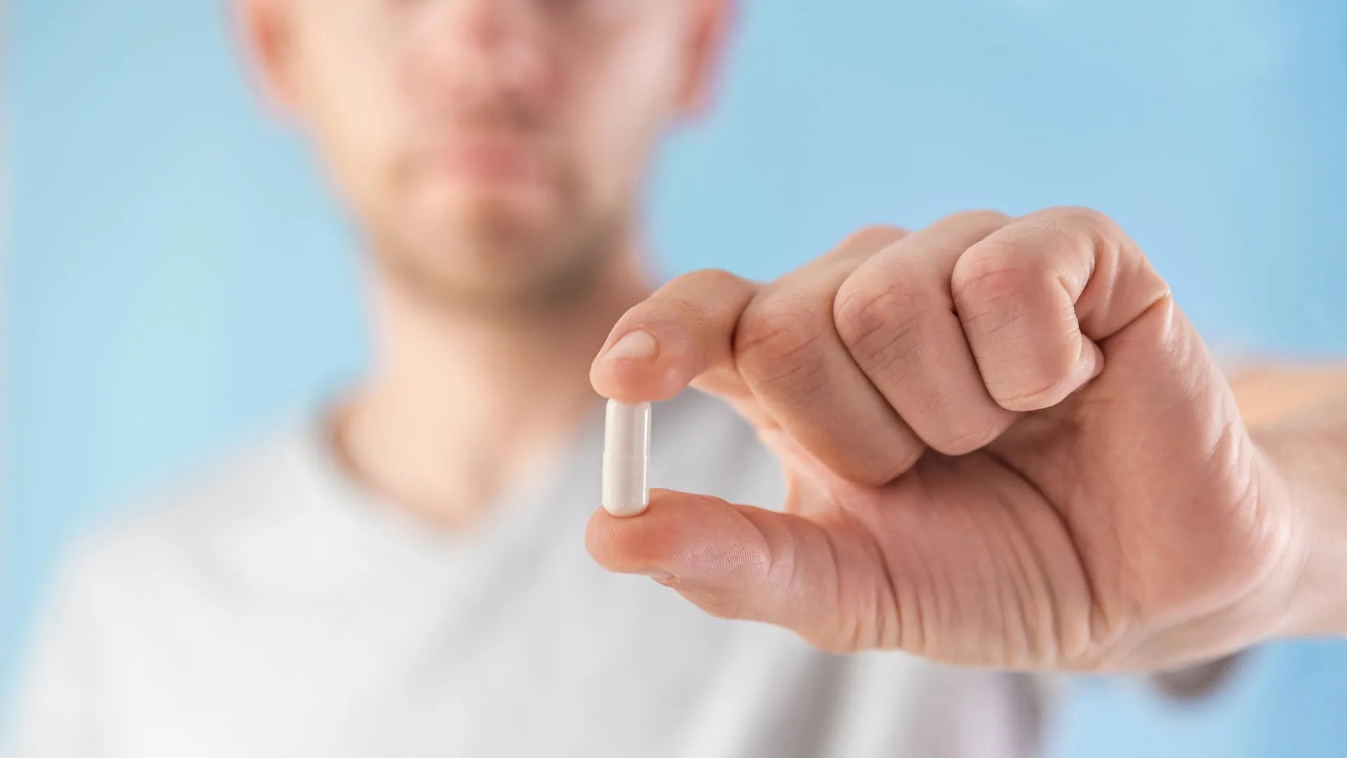
"This discovery is especially meaningful for older individuals and patients who cannot exercise due to frailty, injury, or chronic illness," Dr. Wang noted. "Our findings open the door to developing ‘exercise mimetics’—drugs that chemically activate the Piezo1 pathway to help maintain bone mass and support independence."
The collaborative nature of the project extended across continents, drawing on specialized expertise in pharmacology and cellular biology. Professor Eric Honore, Team Leader at the Institute of Molecular and Cellular Pharmacology within the French National Centre for Scientific Research (CNRS), Université Côte d’Azur (UniCA), and Inserm, who also serves as a Visiting Professor in the Department of Pharmacology and Pharmacy at HKUMed and co-leader of the study, underscored the broader clinical horizon.
"This offers a promising strategy beyond traditional physical therapy," Professor Honore remarked. "In the future, we could potentially provide the biological benefits of exercise through targeted treatments, thereby slowing bone loss in vulnerable groups such as bedridden patients or those with limited mobility, and substantially reducing their risk of fractures."
Chronology and Collaborative Foundations
The publication of these findings in Signal Transduction and Targeted Therapy is the culmination of years of meticulous investigation at the intersection of endocrinology, stem cell biology, and biomechanics. The research project brought together leading minds from HKUMed’s State Key Laboratory of Pharmaceutical Biotechnology and European research infrastructure.
The initiative was sustained by substantial competitive grant funding from multiple regional and international bodies, reflecting the high priority placed on addressing age-related musculoskeletal disorders. Support was mobilized through the Areas of Excellence Scheme and the General Research Fund of the Research Grants Council in Hong Kong; the Health and Medical Research Fund under the Health Bureau of the Government of the HKSAR; the National Key R&D Program of China; the National Natural Science Foundation of China; the Human Frontier Science Program; the French National Research Agency; Fondation de France; Fondation pour la Recherche Médicale; and the Macau Science and Technology Development Fund.
Implications and Future Directions for Clinical Practice
As the research team transitions from basic science discovery to translational drug development, the medical community is closely monitoring the progression toward clinical trials. Developing an effective exercise mimetic requires designing pharmacological compounds that can selectively target Piezo1 on bone marrow stem cells without causing unwanted off-target systemic side effects, given that mechanosensitive channels are also expressed in vascular and neural tissues.
If successful, the clinical application of Piezo1 agonists could fundamentally alter the standard of care for geriatric medicine, orthopedics, and long-term critical care. For patients recovering from severe trauma, individuals paralyzed by spinal cord injuries, or elderly patients confined to prolonged bed rest, the availability of a pharmacological agent that halts bone resorption and stimulates osteogenesis by mimicking mechanical loading would prevent the steep decline in bone density that currently plagues immobilized care.
Ultimately, by bridging the gap between mechanical force and molecular biology, the HKUMed research team has provided a conceptual blueprint for overcoming one of aging biology’s most stubborn challenges. While a pill cannot replace the cardiovascular and metabolic benefits of active lifestyle choices for the general population, the advent of exercise mimetics promises to safeguard the skeletal integrity and personal independence of society’s most vulnerable members.