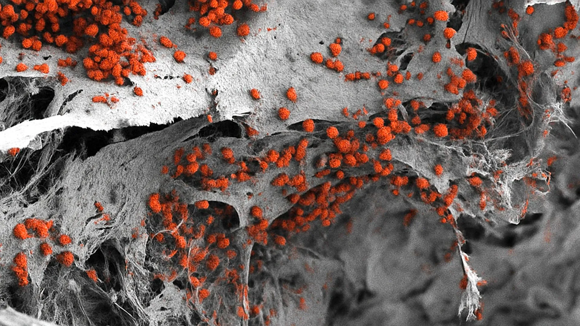
Deep within the skeletal framework of the human body lies one of biology’s most industrious and tightly regulated environments: the bone marrow. Often referred to colloquially as the body’s blood factory, this highly specialized tissue is responsible for generating billions of red blood cells, white blood cells, and platelets every single day to maintain oxygen transport, immune defense, and hemostasis. Operating largely unnoticed under normal physiological conditions, the marrow commands scientific attention only when its complex machinery malfunctions, most notably in devastating hematological malignancies such as leukemias, lymphomas, and multiple myelomas. For decades, however, researchers investigating these conditions faced a formidable bottleneck: the sheer inability to accurately replicate the human bone marrow environment outside of a living organism.
That limitation may soon be a relic of the past. In a landmark achievement published in the esteemed scientific journal Cell Stem Cell, a multidisciplinary team of researchers from the Department of Biomedicine at the University of Basel and University Hospital Basel has successfully recreated the intricate architecture of human bone marrow in a laboratory setting using exclusively human cells. This breakthrough represents a monumental first in the field of biomedical engineering, offering an unprecedented platform that bridges the gap between simplistic cell cultures and complex mammalian models. By providing a faithful 3D simulation of human hematopoiesis—the formation of blood cellular components—this novel system holds profound implications for basic research, pharmacological drug screening, and the burgeoning frontier of personalized medicine, while simultaneously aligning with global mandates to curb the reliance on animal testing in scientific laboratories.
Decoding the Endosteal Niche: The Complexity of Blood Production
To understand the magnitude of the University of Basel team’s achievement, one must appreciate the labyrinthine microenvironment of the bone marrow. The tissue is not a homogenous soup of cells; rather, it is compartmentalized into specialized microenvironments known as niches. Among the most critical of these is the endosteal niche, situated in the immediate vicinity of the inner bone surface. This specific zone plays a pivotal role in regulating hematopoietic stem cells, dictating whether they remain dormant, proliferate, or differentiate into mature blood lineages. Furthermore, the endosteal niche is notoriously implicated in cancer biology, as malignant cells frequently commandeer these microenvironments to evade conventional chemotherapy, leading to relapse and treatment resistance in blood cancer patients.
Replicating the endosteal niche in vitro has long been considered a holy grail of tissue engineering because of its staggering cellular and structural heterogeneity. The niche is a bustling ecosystem comprising not only bone-forming cells such as osteoblasts and osteoclasts, but also an interwoven network of blood vessels, delicate nerve endings, specialized stromal cells, and immune system components. Historically, studying this ecosystem required compromises. Scientists either relied on animal models—predominantly mice—which, despite providing invaluable foundational knowledge, possess significant immunological and physiological divergences from humans, or they utilized two-dimensional cell cultures. These conventional 2D setups fail to capture the mechanical cues, spatial organization, and cell-to-cell signaling dynamics intrinsic to three-dimensional human tissues.
The Chronology of Innovation: From Stem Cells to Functional 3D Architecture
The realization of this human-cell bone marrow model is the culmination of years of rigorous multidisciplinary research in stem cell biology, biomaterials science, and tissue engineering. The project, spearheaded by Professor Ivan Martin and Dr. Andrés García García, required a methodical, step-by-step approach to reconstruct a living tissue matrix from scratch.
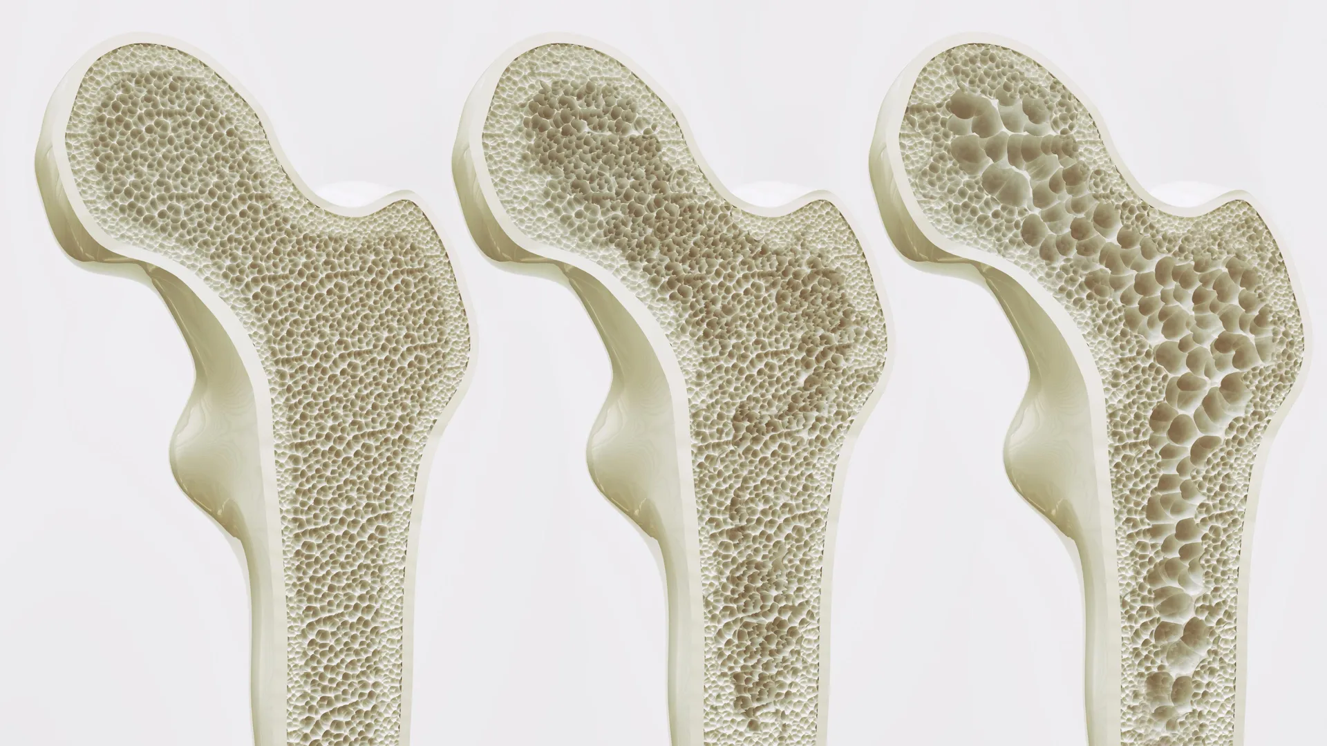
The developmental timeline of the project began with the creation of a biomimetic physical scaffold designed to mimic the natural rigidity and composition of human bone. The researchers utilized hydroxyapatite, a naturally occurring mineral form of calcium apatite that constitutes the major mineral matrix of vertebrate bones and teeth. This porous scaffold provided the necessary structural foundation and mechanical support for subsequent cellular colonization.
Next, the team leveraged cutting-edge molecular biology techniques to reprogram somatic human cells into induced pluripotent stem cells (iPSCs). These versatile stem cells possess the remarkable capacity to differentiate into virtually any cell type found in the human body, driven by the specific biochemical and biophysical cues of their surrounding microenvironment. By introducing these reprogrammed stem cells into the hydroxyapatite scaffold, the researchers initiated a carefully controlled developmental cascade.
Through precise biochemical signaling over a period of weeks, the stem cells were guided to differentiate into the diverse array of specialized cell types that populate natural bone marrow. Subsequent microscopic, biochemical, and genomic analyses revealed that the resulting three-dimensional structure remarkably mirrored the cellular composition, spatial architecture, and functional dynamics of the human endosteal niche. Crucially, the resulting construct achieved a macroscopic scale previously unattained in human-based models, measuring approximately eight millimeters in diameter and four millimeters in thickness. Within this robust 3D system, the researchers successfully maintained human blood cell formation ex vivo for several consecutive weeks, demonstrating sustained biological functionality.
Bridging the Gap: Reducing Animal Experiments in Biomedical Science
Beyond its immediate scientific utility, the creation of this humanized bone marrow model addresses a pressing ethical and regulatory challenge within the global research community: the imperative to reduce, refine, and replace animal experiments—collectively known as the 3Rs principle.
For generations, preclinical research involving hematopoiesis and blood disorders has been overwhelmingly dependent on murine models. While mice have undoubtedly advanced medical science by allowing researchers to observe complex biological processes in a living organism, significant interspecies variations often limit the direct clinical translation of findings from murine studies to human patients. Biological differences in drug metabolism, immune system architecture, and bone marrow microenvironment signaling mean that a treatment proving successful in a mouse model can frequently fail or behave unpredictably in human clinical trials.
Professor Ivan Martin contextualized this paradigm shift, acknowledging the irreplaceable historical contributions of animal models while highlighting the urgent necessity for human-relevant alternatives. "We have learned a great deal about how bone marrow works from mouse studies," Martin noted. "However, our model brings us closer to the biology of the human organism. It could serve as a complement to many animal experiments in the study of blood formation in both healthy and diseased conditions."
By providing a reliable, standardized, and biologically faithful human alternative, the University of Basel platform offers research institutions a viable tool to bypass preliminary animal trials for certain types of investigations. This not only aligns with increasingly stringent institutional and governmental regulations regarding animal welfare in research laboratories but also accelerates the pace of discovery by generating data derived directly from human cellular biology.
Pharmacological Applications and the Challenge of Scalability
While the scientific community has greeted the development with widespread enthusiasm, the research team remains pragmatic about the hurdles that must be cleared before the model can be integrated into high-throughput pharmaceutical pipelines. Drug discovery and toxicology screening typically demand miniaturized, high-throughput assay formats capable of testing dozens of compounds and concentrations simultaneously in rapid succession.
Herein lies the current technological paradox of the Basel model: its greatest physiological strength—its realistic scale and cellular complexity—presents a structural challenge for large-scale drug screening. Measuring nearly a centimeter across, the current iteration of the 3D bone marrow system is too large for conventional high-throughput screening equipment utilized in pharmaceutical laboratories.
Dr. Andrés García García addressed this operational bottleneck, outlining the engineering steps required for industrial translation. "However, for this specific purpose, the size of our bone marrow model might be too large," García García explained. To adapt the platform for widespread pharmacological screening, the research group is actively exploring methods to miniaturize the system without sacrificing the intricate cellular interactions and spatial organization that make the current model biologically authentic. Achieving this balance of scale and complexity will be a primary focus of subsequent engineering phases.
The Horizon of Personalized Medicine in Hematology
Perhaps the most visionary implication of the University of Basel’s breakthrough lies in the realm of personalized medicine. Treating hematological malignancies such as acute myeloid leukemia or multiple myeloma remains exceptionally challenging due to high inter-patient variability and the tendency of cancer cells to develop resistance to standard chemotherapeutic regimens.
Looking toward the future, the research team envisions a clinical paradigm where patient-specific bone marrow models could be synthesized in the laboratory. By taking a small tissue biopsy or skin sample from an individual patient, reprogramming their cells into iPSCs, and constructing a personalized 3D bone marrow replica, oncologists could directly test a panel of different drugs and therapeutic combinations on the patient’s own living cells ex vivo.
This bespoke approach would allow physicians to empirically identify the most efficacious treatment strategy for a specific patient prior to administering potentially toxic chemotherapy regimens, thereby maximizing therapeutic efficacy while minimizing adverse side effects. Although realizing this clinical vision will necessitate substantial further optimization, standardization, and cost-reduction efforts, the successful construction of a functional human-cell bone marrow model marks a critical foundational stride.
Broader Implications for Translational Medicine
The successful recreation of the human bone marrow niche in vitro signifies a watershed moment in tissue engineering and translational hematology. By transcending the limitations of two-dimensional cultures and reducing structural reliance on animal models, the University of Basel team has unlocked a sophisticated window into the human hematopoietic system.
As the researchers refine the platform for pharmaceutical scalability and advance toward patient-specific applications, the scientific community stands on the threshold of a new era in blood disorder research. With enhanced capabilities to study leukemic drug resistance, screen novel pharmacological agents on human tissue, and map the subtle molecular dialogues within the endosteal niche, this artificial blood factory promises to reshape the landscape of modern medicine, bringing safer therapies and personalized treatments ever closer to clinical reality.