Researchers at the Indiana University School of Medicine have successfully engineered an advanced imaging methodology designed to visualize the intricate cellular landscape of mouse bone marrow. Published recently in the peer-reviewed journal Leukemia, this scientific breakthrough overcomes long-standing biological and technical hurdles associated with examining gelatinous soft tissue encased within rigid skeletal structures. By harnessing the capabilities of a sophisticated multiplex platform known as Phenocycler 2.0, the multidisciplinary research team has opened new pathways for investigating complex hematological disorders, oncological diseases, and autoimmune conditions.
The successful adaptation of this technology provides the scientific community with an unprecedented capability to map dozens of cellular markers simultaneously within structurally intact tissue. This innovation is expected to significantly accelerate the pipeline for novel therapeutics, offering researchers a high-resolution window into the primary site of hematopoiesis—the biological process responsible for the continuous generation of blood and immune cells.
Navigating the Historical Challenges of Bone Marrow Analysis
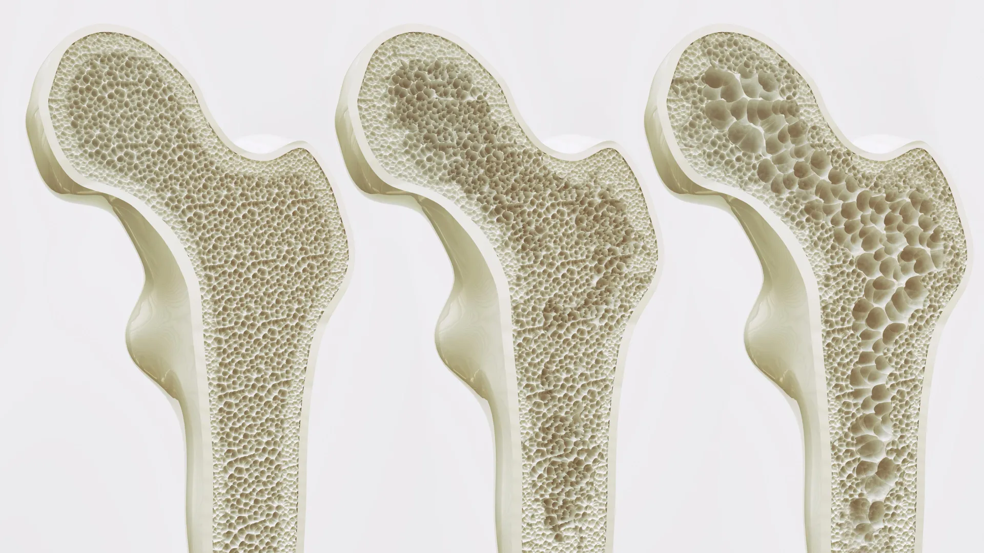
For decades, the study of bone marrow has presented a formidable challenge to biomedical researchers. Anatomically, bone marrow occupies a uniquely difficult niche: it is a soft, highly dynamic, and gelatinous tissue securely protected by dense cortical and trabecular bone. This duality has historically forced scientists to compromise when attempting to analyze its cellular composition.
Prior to this advancement, the scientific community relied heavily on traditional modalities such as flow cytometry and standard fluorescence microscopy. While these methods have historically driven major discoveries in immunology and hematology, they carry inherent limitations. Flow cytometry, though powerful for quantifying distinct cell populations, requires the mechanical or enzymatic dissociation of the tissue. This destructive process obliterates the spatial architecture of the bone marrow, stripping researchers of vital context regarding how different cell types are spatially organized relative to one another and their surrounding microenvironment.
Conversely, standard fluorescence imaging preserves tissue architecture but suffers from severe multiplexing constraints. Traditional fluorescence microscopy is typically limited to visualizing only three to four cellular markers simultaneously due to spectral overlap among fluorophores. Consequently, researchers were previously forced to stitch together a fragmented understanding of bone marrow biology by analyzing multiple serial sections, introducing variability and missing the holistic interactions occurring within the native microenvironment.
Recognizing these barriers, the Indiana University team sought to adapt the Phenocycler 2.0 platform—a technology previously validated for use in solid organs such as the spleen and kidneys—to conquer the unique structural hurdles posed by murine skeletal systems.
Chronology of the Research and Methodological Breakthrough
The journey toward this methodological milestone required extensive optimization, interdisciplinary collaboration, and rigorous validation by the IU Cooperative Center of Excellence in Hematology.
Initial phases of the project began as researchers sought to address the difficulties of preparing bone marrow samples without degrading delicate cellular epitopes or damaging the surrounding mineralized matrix. By refining tissue decalcification, sectioning, and cyclic staining protocols, the research team successfully integrated the Phenocycler 2.0 system into their skeletal biology workflow.
Unlike traditional methods, the Phenocycler 2.0 utilizes an automated iterative staining and imaging cycle powered by DNA-barcoded antibodies. This cyclical process allows researchers to apply a panel of antibodies, image the targets, gently cleave the fluorescent tags, and repeat the process with a fresh set of markers on the exact same tissue section.
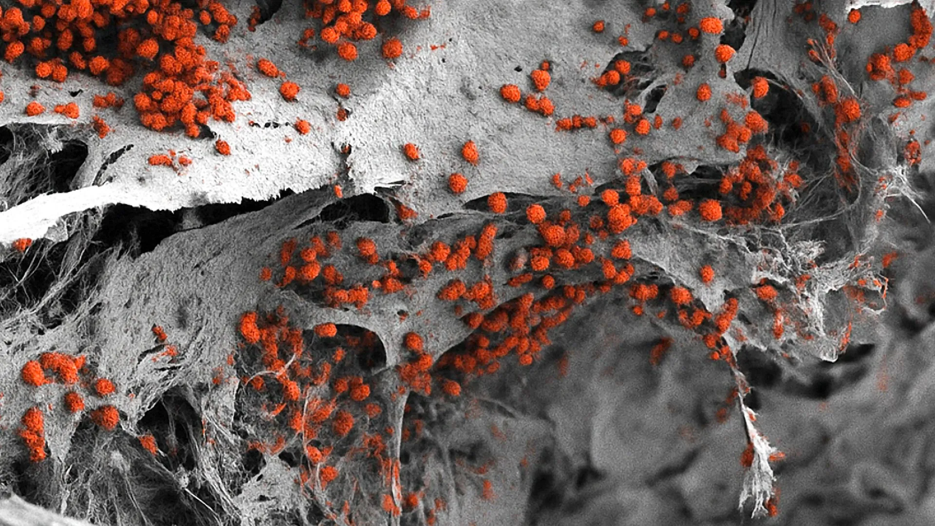
In their landmark study, the IU team achieved a historic milestone by successfully visualizing 25 distinct cellular markers within a single, intact section of mouse bone marrow. This leap from three markers to 25 represents an exponential increase in data density, allowing scientists to map out rare stem cell niches, immune cell lineages, vascular networks, and stromal components with subcellular resolution.
Following the successful generation and validation of these high-dimensional imaging datasets, the findings underwent peer review and were accepted for publication in Leukemia. Concurrently, recognizing the profound commercial and clinical potential of the optimized protocol, the Indiana University Innovation and Commercialization Office filed a provisional patent for the novel imaging methodology to protect the intellectual property and pave the way for future translational applications.
Supporting Data and Technical Scope
The implications of the newly developed technique are deeply rooted in the richness of the data it generates. Bone marrow is not a homogenous soup of cells; rather, it is a highly compartmentalized organ where hematopoietic stem cells (HSCs) reside in specialized niches that dictate whether they remain dormant, self-renew, or differentiate into red blood cells, platelets, or various white blood cells.
By capturing 25 markers simultaneously in situ, researchers can now identify rare cell populations in their exact physiological neighborhoods. This includes charting the spatial distribution of hematopoietic stem and progenitor cells relative to endosteal surfaces, sinusoidal blood vessels, sympathetic nerve fibers, and immunoregulatory cells such as regulatory T cells and macrophages.
The research team was spearheaded by co-lead author Sonali Karnik, PhD, an assistant research professor of orthopedic surgery at the IU School of Medicine, alongside co-senior author Reuben Kapur, PhD, director of the Herman B Wells Center for Pediatric Research and co-director of the IU Cooperative Center of Excellence in Hematology.
The broader research group comprised a robust coalition of IU investigators, including Connor Gulbronson, Paige C. Jordan, Rahul Kanumuri, Baskar Ramdas, Ramesh Kumar, Melissa L. Hartman, Izza Khurram, Drew M. Brown, Karen E. Pollok, Pratibha Singh, and Melissa A. Kacena. Financial backing for the multi-year investigative effort was provided by the National Institutes of Health, underscoring the federal significance of advancing fundamental tools for hematological and oncological research.
Official Perspectives and Expert Analysis
The scientific leadership at Indiana University School of Medicine has emphasized the transformative nature of the technology, highlighting its broad applicability across various branches of biomedical science.
"Bone marrow is difficult to study because it is gelatinous and encased in hard bone," explained Dr. Karnik. "Since bone marrow plays an important role in blood and immune cell formation and houses valuable stem cells, our unique imaging approach offers a useful tool for a variety of research applications."
Because murine models serve as the foundational bedrock for preclinical trials and human disease modeling worldwide, the ability to visualize the murine bone marrow microenvironment with such granular detail addresses a major industry bottleneck.
"Because mouse models are widely used to study human diseases, this technique offers a promising new method for investigating a range of conditions like autoimmune diseases, leukemia and other disorders involving bone marrow," noted Dr. Kapur.
Experts outside the immediate research group have noted that spatial biology is currently revolutionizing oncology and immunology. While spatial transcriptomics and multiplex imaging have transformed the study of solid tumors—such as breast, lung, and colon cancers—bone marrow malignancies and non-malignant bone marrow disorders have historically lagged behind due to the aforementioned technical barriers. By bridging this gap, the IU team’s methodology positions bone marrow research at the cutting edge of spatial multi-omics.
Broader Impact, Clinical Implications, and Future Directions
The implications of this breakthrough extend far beyond basic academic discovery, holding profound promise for translational medicine, drug development, and targeted therapeutics.
Advancing Oncology and Leukemia Research
Leukemias, multiple myelomas, and myelodysplastic syndromes originate within the bone marrow microenvironment. Often, malignant cells hijack normal stem cell niches, evading chemotherapeutic agents and fostering drug resistance. With the ability to visualize 25 or more markers simultaneously in intact tissue, researchers can now observe how leukemic cells alter the spatial organization of normal hematopoietic stem cells, how they interact with blood vessels, and how they suppress immune surveillance within the bone marrow niche. This granular insight could lead to the identification of novel therapeutic targets designed to disrupt the protective microenvironments that shield cancer cells from treatment.
Understanding Autoimmune Diseases
Many autoimmune disorders—including lupus, rheumatoid arthritis, and various systemic inflammatory conditions—involve aberrations in immune cell development and central tolerance, processes heavily governed by the bone marrow and secondary lymphoid organs. The new imaging technique allows immunologists to track autoreactive clones and inflammatory cytokine networks within their native anatomical context, potentially revealing new biomarkers and intervention strategies.
Musculoskeletal Disorders and Regenerative Medicine
Because the research originated in the Department of Orthopedic Surgery, the technique also holds immense value for musculoskeletal science. Bone marrow interfaces directly with bone tissue, playing a critical role in bone remodeling, fracture healing, and skeletal homeostasis. Understanding the crosstalk between skeletal stem cells, osteoblasts, osteoclasts, and hematopoietic elements within the marrow cavity can illuminate pathways for treating osteoporosis, osteogenesis imperfecta, and degenerative joint diseases.
Expanding the Technological Horizon
Building upon their initial success, the Indiana University research team is not resting on its laurels. Current developmental efforts are actively focused on expanding the marker panel. Future iterations of the methodology aim to incorporate structural and physiological features that extend beyond standard protein markers, including specialized markers for bone matrix components, peripheral nerves, smooth muscle, and an even broader array of immune and signaling cell phenotypes.
As the provisional patent process moves forward through the IU Innovation and Commercialization Office, the scientific community anticipates that this methodology will be widely adopted by academic research centers and pharmaceutical companies alike. By turning a previously opaque and inaccessible tissue into an open book of high-resolution spatial data, Indiana University School of Medicine has established a powerful new paradigm in the ongoing quest to understand, treat, and ultimately cure complex diseases of the blood and bone.