Led by associate professor of developmental biology Kyle Loh, the research team discovered that the human brain is actually a composite structure, formed by the convergence of two ancient, independent nervous systems. This finding not only rewrites the textbook understanding of human anatomy but also solves a persistent technical bottleneck that has hindered medical research into neurodegenerative diseases for decades.
The Anatomy of a Dual Origin
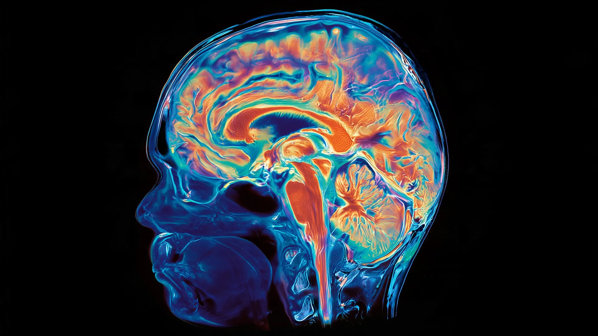
The human brain is traditionally categorized into three functional tiers: the forebrain, the midbrain, and the hindbrain. The forebrain serves as the epicenter of complex cognition, facilitating the higher-order functions that define human existence, including linguistic processing, abstract mathematics, self-reflection, and consciousness. Conversely, the hindbrain—often synonymous with the brain stem—functions as the biological autopilot. It is the site of vital, life-sustaining regulation, managing respiratory rhythms, heart rate, sleep cycles, and caloric intake. Furthermore, it orchestrates the intricate motor coordination required for speech and deglutition, or swallowing.
The research conducted at Stanford demonstrates that these two regions are not merely different sectors of the same developmental tree; they are distinct entities with entirely different "birth certificates." By examining mouse embryos during the critical stage of gastrulation—the period when an organism’s basic body plan is established—the researchers identified two separate progenitor cell populations.
The forebrain and midbrain originate from cells expressing the gene Otx2. In contrast, the hindbrain arises from cells marked by the expression of the Gbx2 gene. This segregation is not a localized occurrence but is hardcoded into the cell’s architecture from the earliest moments of embryonic development. The team observed that the chromatin—the complex of DNA and proteins that dictates gene accessibility—is configured fundamentally differently in the anterior versus the posterior neural ectoderm. These divergent epigenetic landscapes effectively commit these cell populations to independent developmental pathways, meaning the hindbrain does not "evolve" from the forebrain, nor vice versa.
Historical Context and the Laboratory Hurdle
The difficulty of replicating this process in the laboratory has been one of the most frustrating barriers in clinical neuroscience. For years, researchers attempting to generate hindbrain neurons from human pluripotent stem cells struggled to achieve consistent results. The Stanford study suggests that these failure rates were not due to lack of skill or proper chemical signaling, but rather a fundamental misunderstanding of cellular lineage.
Previous protocols attempted to "coax" cells meant for forebrain development into adopting a hindbrain fate. According to the research, this is biologically impossible. Because the hindbrain follows a distinct developmental trajectory from the moment of inception, trying to force a forebrain progenitor into a hindbrain role is akin to trying to turn a blueprint for a skyscraper into the structural foundation for a submarine. By recognizing this split, the researchers were able to successfully guide human stem cells to differentiate specifically into functional hindbrain motor neurons for the first time. These laboratory-grown cells displayed authentic physiological markers, including the generation of electrical action potentials and the synthesis of proteins required for facial and swallowing motor control.
Evolutionary Timeline: A 500-Million-Year Inheritance
To contextualize these findings, the team conducted a comparative analysis of evolutionary biology spanning over half a billion years. They discovered that this dual-system architecture is not a recent innovation of primates or even mammals; it is an ancient heritage. Evidence of this two-origin arrangement was identified in chickens, zebrafish, and even the humble acorn worm, which shares a deep evolutionary lineage with humans.
Tracing the timeline back further, the researchers noted that jellyfish—organisms that diverged from the human lineage approximately 600 to 700 million years ago—possess two separate neural networks located at different poles of their bodies. The implication is that the vertebrate brain is a product of evolutionary "bricolage," where two separate, primordial nervous systems were physically brought together over hundreds of millions of years. This discovery challenges the notion of the brain as a perfectly integrated organ, suggesting instead that it is an efficient, albeit evolutionary, compromise that preserved the autonomy of its two constituent parts.
Clinical Implications for ALS and SMA
The immediate impact of this discovery lies in the realm of clinical pathology. Spinal muscular atrophy (SMA) and amyotrophic lateral sclerosis (ALS) are debilitating conditions characterized by the progressive degradation of motor neurons, many of which reside in the hindbrain. SMA is a leading genetic cause of mortality in infants, while ALS typically manifests in middle age, causing a devastating loss of muscle control that eventually compromises the ability to swallow and breathe.
Because the hindbrain is shielded by the skull and inaccessible for routine biopsy, clinical researchers have been forced to rely on indirect methods or animal models that do not perfectly replicate human pathology. The ability to generate authentic human hindbrain neurons in a petri dish provides a novel, high-fidelity model for drug testing and disease modeling. Scientists can now observe the cellular progression of ALS and SMA in real-time, testing how specific mutations impact the survival of these unique hindbrain neurons.
Furthermore, this research has potential downstream applications for metabolic medicine. Given that the hindbrain contains the neural circuitry responsible for regulating hunger, the ability to model these cells in vitro could lead to a deeper understanding of how weight-loss therapeutics, such as GLP-1 receptor agonists like semaglutide, interact with the brain’s hunger-regulation centers.
The Future of Regenerative Neuroscience
The study, co-authored by graduate students Carolyn Dundes and Rayyan Jokhai, marks a departure from the "neuron-first" focus of modern stem cell biology. By shifting the attention back to the earliest stages of embryonic development, the team has unlocked a new paradigm for regenerative medicine. The research suggests that the future of treating neurodegenerative conditions does not lie solely in the repair of mature neurons, but in understanding the distinct developmental "instructions" that give rise to specific brain regions.
"We have shown for the first time that the front of the brain arises from a totally different progenitor cell than the back of the brain," Loh stated. This assertion is supported by an extensive list of funding bodies, including the National Institutes of Health, the National Science Foundation, and the California Institute for Regenerative Medicine, all of which underscore the high degree of scientific confidence in the team’s findings.
As the scientific community begins to digest the implications of this dual-origin brain, the focus will likely turn to the spinal cord and other neural structures that may also follow independent developmental paths. By mapping the "architectural blueprints" of the central nervous system, researchers are moving closer to a future where developmental biology can be leveraged to address some of the most intractable diseases in human medicine. The brain, once thought to be a singular, monolithic organ, is now revealed as a complex, evolutionary hybrid—a discovery that promises to reshape neurology for the next generation.