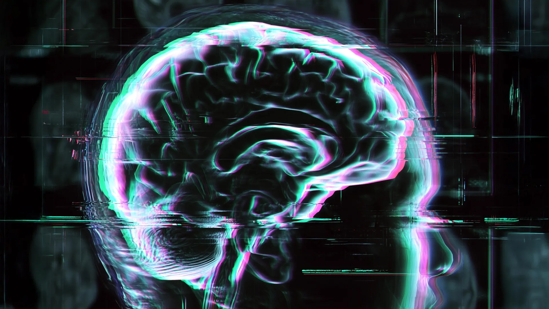
A groundbreaking study published in the open-access journal PLOS Medicine has provided significant new insights into how various neurological, psychiatric, and addictive disorders manifest as "accelerated aging" within the human brain. Led by Shile Qi of the Nanjing University of Aeronautics and Astronautics in China, the research utilized advanced neuroimaging techniques to determine that conditions ranging from Alzheimer’s disease to alcohol addiction leave unique biological signatures on the brain’s structure, effectively causing it to appear chronologically older than it is.
The research team analyzed structural magnetic resonance imaging (MRI) data from a massive cohort of 48,598 individuals. By comparing 45,900 healthy controls against 2,698 patients diagnosed with a spectrum of conditions—including Alzheimer’s disease (AD), mild cognitive impairment (MCI), schizophrenia, bipolar disorder, major depressive disorder, attention-deficit/hyperactivity disorder (ADHD), autism spectrum disorder (ASD), and substance addictions—the researchers were able to quantify the discrepancy between an individual’s chronological age and their "brain age."
The Mechanics of Predictive Age Difference
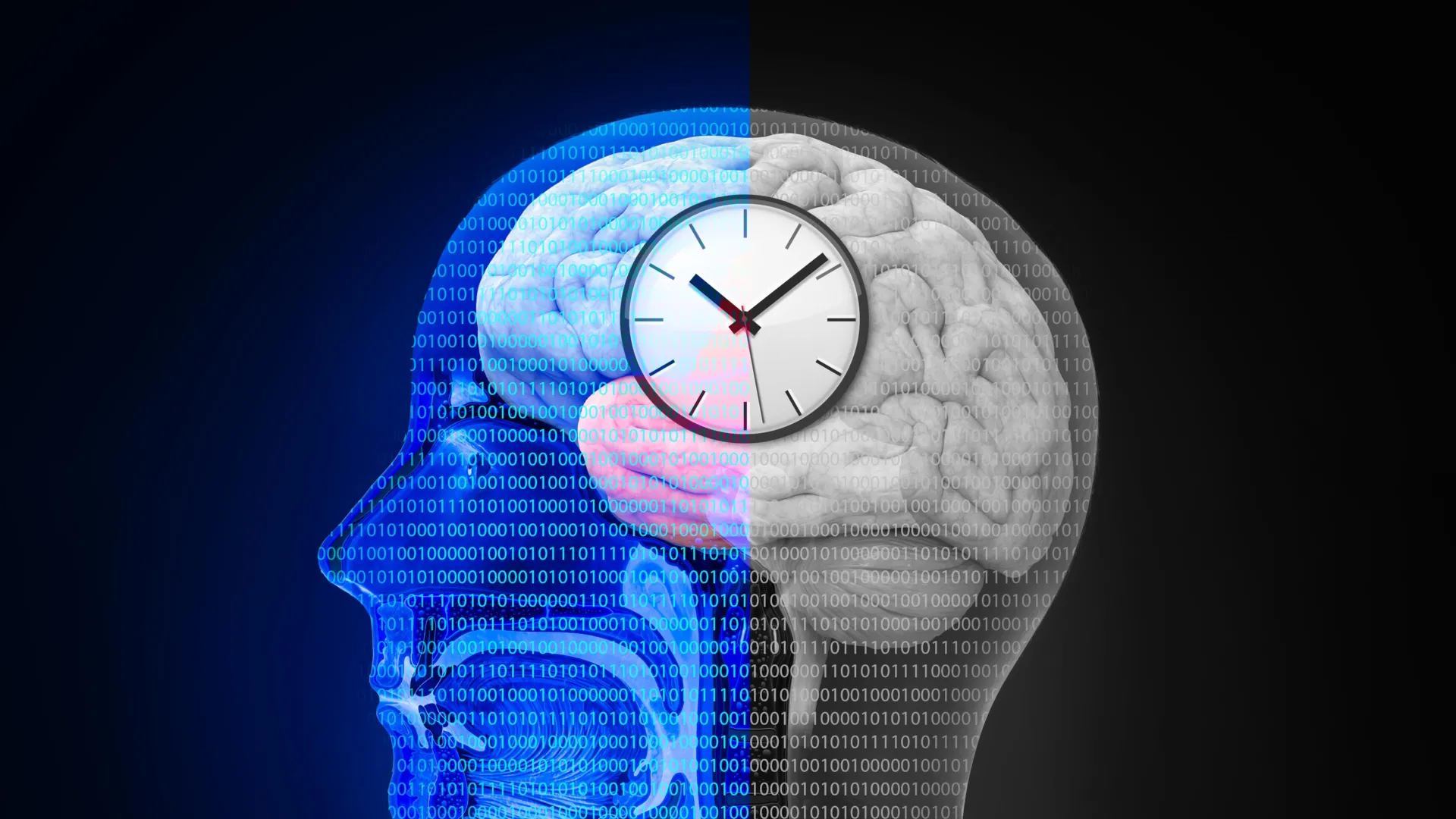
At the heart of this study is the metric known as Predictive Age Difference (PAD). This diagnostic tool allows researchers to calculate a "brain age" based on structural MRI scans—which measure grey matter volume, cortical thickness, and other morphological indicators—and compare it against the patient’s actual chronological age. A positive PAD value indicates that a subject’s brain exhibits the physical characteristics of an older organ, suggesting a premature decline or biological weathering.
While the concept of "brain age" has been explored in prior literature, this study distinguishes itself through its breadth and granular mapping. Rather than viewing accelerated aging as a monolithic phenomenon, the authors argue that the brain ages differently depending on the specific underlying pathology. This suggests that the biological "clocks" for different mental health conditions are regulated by distinct neural pathways and gene expression profiles.
Chronology of Brain Aging Research
The quest to quantify brain aging through imaging is a relatively recent development in neuroscience, gaining momentum over the last decade. Historically, scientists relied on post-mortem examinations to study the physical degradation of the brain. The advent of high-resolution structural MRI in the early 2000s allowed for non-invasive longitudinal studies.
By 2015, researchers began utilizing machine learning algorithms to map the normative aging process, establishing a baseline for what a "healthy" brain should look like at ages 20, 40, 60, and beyond. This latest study by Qi et al. represents the culmination of several years of data aggregation, drawing from multiple international brain imaging databases. It marks a shift from simply observing decline to categorizing the specific regional patterns of that decline across a wide variety of psychiatric and neurological diagnostic categories.
Findings: Neurodegeneration vs. Psychiatric Disorders
The data yielded stark contrasts between categories of disorders. As anticipated, neurodegenerative conditions such as Alzheimer’s disease and mild cognitive impairment displayed the most significant positive PAD values. This confirms that these conditions act as catalysts for rapid structural loss, particularly in areas associated with memory, executive function, and sensory processing.
However, the study also revealed that addiction and psychiatric disorders contribute to significant, albeit different, manifestations of brain aging. The research highlighted that while individuals with schizophrenia and major depressive disorder showed increased PAD, the patterns were localized differently than those found in dementia patients.
Perhaps most surprising was the discovery of what the brain did not show. The study found no statistically significant difference in PAD between healthy controls and individuals diagnosed with ADHD or ASD. This finding suggests that while these neurodevelopmental conditions involve clear functional differences, they may not necessarily accelerate the structural aging process in the same way that progressive psychiatric or degenerative disorders do.
Mapping the Brain: Regional Vulnerabilities
A primary contribution of the Nanjing University study is its regional breakdown of brain aging. By examining individual brain regions, the researchers mapped how specific disorders "sculpt" the aging process:
- The Prefrontal Cortex: This region, essential for decision-making and personality expression, showed elevated PAD across a broad spectrum of disorders. Its sensitivity makes it a primary site for the systemic impact of mental health conditions.
- Frontal and Temporal Lobes: These areas showed higher PAD specifically in patients with psychiatric disorders, potentially explaining the cognitive and emotional volatility often associated with these conditions.
- The Default Mode Network and Thalamus: These regions were heavily affected in cases of addiction. The default mode network, which is active during wakeful rest and self-referential thought, appears particularly vulnerable to the structural changes associated with substance dependency.
- Occipital Cortex: Linked primarily with dementia, the aging of this visual processing area suggests that the physical toll of neurodegeneration is more pervasive than previously categorized in localized mapping.
Scientific Implications and Biomarker Development
The correlation between these brain aging patterns and gene expression suggests that the phenomenon is rooted in biology rather than merely being a byproduct of lifestyle or stress. The researchers identified specific patterns of gene transcription associated with the observed aging, indicating that different conditions trigger distinct molecular pathways.
"Different neurological disorders appear to leave different signatures on the brain aging clock," the study authors noted. This assertion has significant implications for clinical practice. If these "signatures" can be reliably mapped, they could eventually serve as biomarkers—biological indicators that allow clinicians to diagnose conditions earlier, track the progression of a disease, or even assess how a patient is responding to a particular therapy.
Contextualizing the Limitations
While the findings are robust, the research team emphasizes that this study is correlational. It does not definitively prove causation; it is not yet clear whether the accelerated aging is a cause of the psychiatric condition or a result of it. Furthermore, the researchers acknowledge the difficulty of "comorbidity." Many individuals with psychiatric disorders also struggle with substance addiction, and these overlapping conditions can obscure the specific structural impact of any single diagnosis.
External experts in the field of geriatric psychiatry have noted that while the study is a milestone, it must be interpreted within the context of systemic health. Factors such as chronic inflammation, cardiovascular health, and socioeconomic stress are known to influence brain aging, and these variables can be difficult to fully isolate in large-scale imaging databases.
Future Directions for Neuro-Aging Research
The implications for the medical community are profound. By moving toward a model where brain disorders are viewed through the lens of structural aging, researchers may be able to develop interventions that target the specific biological pathways identified in the study. If the brain’s "aging clock" can be slowed or stabilized, the severity of symptoms for conditions like schizophrenia or addiction could potentially be mitigated.
As data collection continues, the next phase of research will likely focus on longitudinal studies—tracking the same individuals over decades to see how the PAD value changes as their condition evolves. This would shift the current static snapshot of brain health into a dynamic model of disease progression.
For now, the study provides a vital roadmap for future investigation. By distinguishing the aging signatures of the prefrontal cortex from those of the thalamus, and linking these to specific diagnostic categories, Shile Qi and his team have provided a new framework for understanding the profound physical toll that mental and neurological disorders exact on the human brain. The research serves as a reminder that the brain is not merely a collection of circuits, but a biological organ susceptible to the temporal pressures of the conditions it hosts.