The fundamental challenge of biological life lies in how negatively charged DNA molecules, which naturally repel one another due to their shared electrical polarity, manage to find, recognize, and pair with matching sequences inside the crowded environment of a living cell. This phenomenon, essential for genetic recombination, DNA repair, and gene silencing, has long puzzled researchers. New research published in the journal Nucleic Acids Research has finally provided a definitive, high-resolution view of how this occurs. By utilizing atomic force microscopy and sophisticated molecular dynamic simulations, a multi-institutional research team has validated the long-standing "DNA zipper" model, revealing that positively charged metal ions act as the essential bridge that overcomes electrostatic repulsion to allow for precise genetic alignment.
A Two-Decade Quest for the DNA Zipper
The history of this discovery dates back approximately twenty years, when Professor Alexey Kornyshev of Imperial College London and his colleagues first proposed a theoretical framework for DNA-DNA recognition. Their "DNA zipper" model posited that the salt ions naturally present in the cellular environment might do more than simply shield electrical charge; they might, in fact, organize themselves into specific, repeating patterns. According to this hypothesis, these ion patterns would alter the electrical landscape of the DNA double helix, allowing neighboring molecules to align groove-to-groove, much like the interlocking teeth of a zipper or two spiral staircases meeting in perfect synchronization.
For nearly two decades, this remained a compelling, albeit unproven, hypothesis. The primary obstacle was the sheer scale of the interaction. DNA molecules are nanometers in width, and the ions mediating their interaction are sub-nanometer, making real-time, direct observation of the process nearly impossible with traditional laboratory equipment. The breakthrough required a convergence of advanced physical imaging and computational power, a milestone finally achieved by researchers from the University of York, the University of Sheffield, and Imperial College London.
The Synergy of Microscopy and Simulation
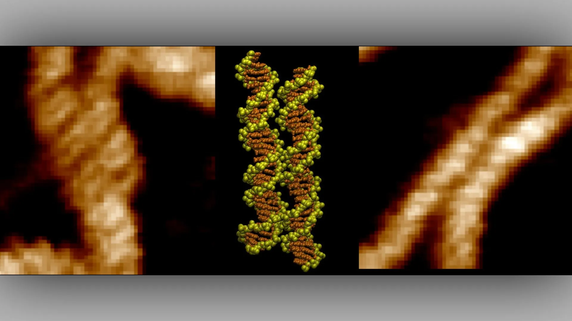
The research team employed a dual-track methodology to bridge the gap between theory and observation. First, they utilized atomic force microscopy (AFM), a high-resolution scanning technique that uses a physical probe to map the topography of surfaces at the molecular level. By scanning DNA samples, the team created detailed topographical maps that provided direct visual evidence of DNA molecules aligning with extraordinary precision. These maps demonstrated that DNA segments did not merely approach one another randomly; they oriented themselves to match their major and minor grooves with a high degree of geometrical fidelity.
However, microscopy alone cannot explain the "why" behind the movement. To understand the underlying physical forces, the team utilized advanced computer simulations that tracked the behavior of individual atoms and ions within the DNA environment. These simulations allowed researchers to visualize how divalent metal ions—positively charged atoms—behaved in the space between the two DNA strands.
The data revealed that these ions do not simply float passively; they function as active molecular agents. A double-charged ion, the simulations showed, can effectively serve as a "bridge" with two charged arms. Each arm interacts with a different DNA molecule simultaneously, creating an attractive force that counteracts the natural repulsion of the negatively charged phosphate backbones. By anchoring themselves into the grooves of the DNA, these ions stabilize the complex, holding the two strands in a locked, parallel alignment that is stable enough to facilitate further biological processes.
Official Responses and Scientific Significance
The significance of this discovery extends beyond mere fundamental physics. Dr. Thomas Catley, co-lead author from the School of Chemical Materials and Biological Engineering at the University of Sheffield, emphasized the rarity of observing such long-hypothesized mechanisms in real-time. "It was incredible to be able to directly visualize the long-hypothesized mechanism for the first time," Dr. Catley noted. "The advanced imaging techniques at our disposal are allowing us to uncover these key DNA interactions which have implications in many key cellular processes."
Dr. Victor Velasco-Berrelleza, who spearheaded the computational modeling efforts, highlighted the necessity of the interdisciplinary approach. "Microscopy shows us what happens, but the simulations allow us to uncover the molecular mechanism behind it," he stated. This sentiment is echoed by Professor Agnes Noy of the University of York, who co-led the research. According to Professor Noy, the discovery provides a critical roadmap for identifying specific genomic regions that are prone to pairing. "This discovery could help researchers identify regions of the genome specially involved in DNA pairing," Noy explained. "These regions may become particularly important when mutations disrupt normal cellular processes and contribute to cancer."
Genomic Hotspots and the Implications for Disease
One of the most provocative findings in the study is the discovery that DNA does not pair with equal affinity across all sequences. The researchers identified specific "hotspots" where the pairing mechanism is significantly stronger. This indicates that certain genetic sequences are biologically programmed to interact more readily than others.
From a clinical perspective, this is a pivotal realization. In the context of cancer, mutations can disrupt the delicate balance of gene regulation. If specific DNA sequences are "programmed" to pair or recognize each other, a mutation that alters the structure of these sequences—or the density of the ions surrounding them—could lead to genomic instability. By mapping these hotspots, scientists may eventually be able to predict which areas of the genome are most susceptible to recombination errors, providing a new dimension to our understanding of oncogenesis and the development of targeted therapies.
Broader Impact: From Biotechnology to Synthetic Biology
While the immediate applications of this research are grounded in cell biology and oncology, the potential for biotechnology is vast. The ability of DNA to self-assemble via ion-mediated bridges offers a blueprint for "DNA nanotechnology." In this field, researchers design synthetic DNA structures for use in drug delivery, biosensing, and molecular computing.
Current methods of DNA assembly often rely on base-pairing rules (adenine to thymine, cytosine to guanine). By understanding and leveraging the ion-mediated "zipper" effect, engineers could theoretically design new classes of DNA structures that assemble based on electrostatic "hotspots" rather than just sequence complementarity. This could allow for the creation of more robust, complex, and programmable nanomaterials that can operate within the harsh, high-salt environments of the human body, where traditional DNA structures might otherwise fail.
Future Directions and Conclusion
The publication of "Imaging and mechanism of DNA-DNA recognition mediated by divalent ions" marks the end of a long period of theoretical speculation and the beginning of a new era of experimental validation. The scientific community now possesses both the visual proof and the physical model required to understand how the architecture of life is maintained at the atomic level.
Future research will likely focus on the role of different types of ions and the effect of varying cellular conditions on the strength of these DNA bridges. For instance, how do changes in cellular pH or the presence of various proteins affect the "zipper" mechanism? As researchers begin to answer these questions, the ability to manipulate these interactions could become a cornerstone of regenerative medicine and genetic engineering. By mastering the molecular bridges that hold our genetic code together, science moves one step closer to correcting the errors that lead to disease and engineering the materials that will define the next generation of medical technology. The "DNA zipper" is no longer just a model; it is a observable, measurable, and potentially exploitable reality of the biological world.