
A single molecular switch is essential for blood stem cells to enter an activated, regenerative state in which they produce new blood cells, according to a preclinical study led by Weill Cornell Medicine investigators. This discovery, published on February 25 in Nature Immunology, represents a significant leap forward in regenerative medicine, offering a potential roadmap for increasing the success rates of bone marrow transplants and enhancing the efficacy of gene therapies for debilitating blood disorders. By identifying the protein FLI-1 as the primary regulator of blood stem cell mobilization, researchers have unlocked a method to "wake up" these cells from their natural state of dormancy, allowing them to multiply and engraft more effectively into patients.
The Biological Mechanics of Stem Cell Quiescence
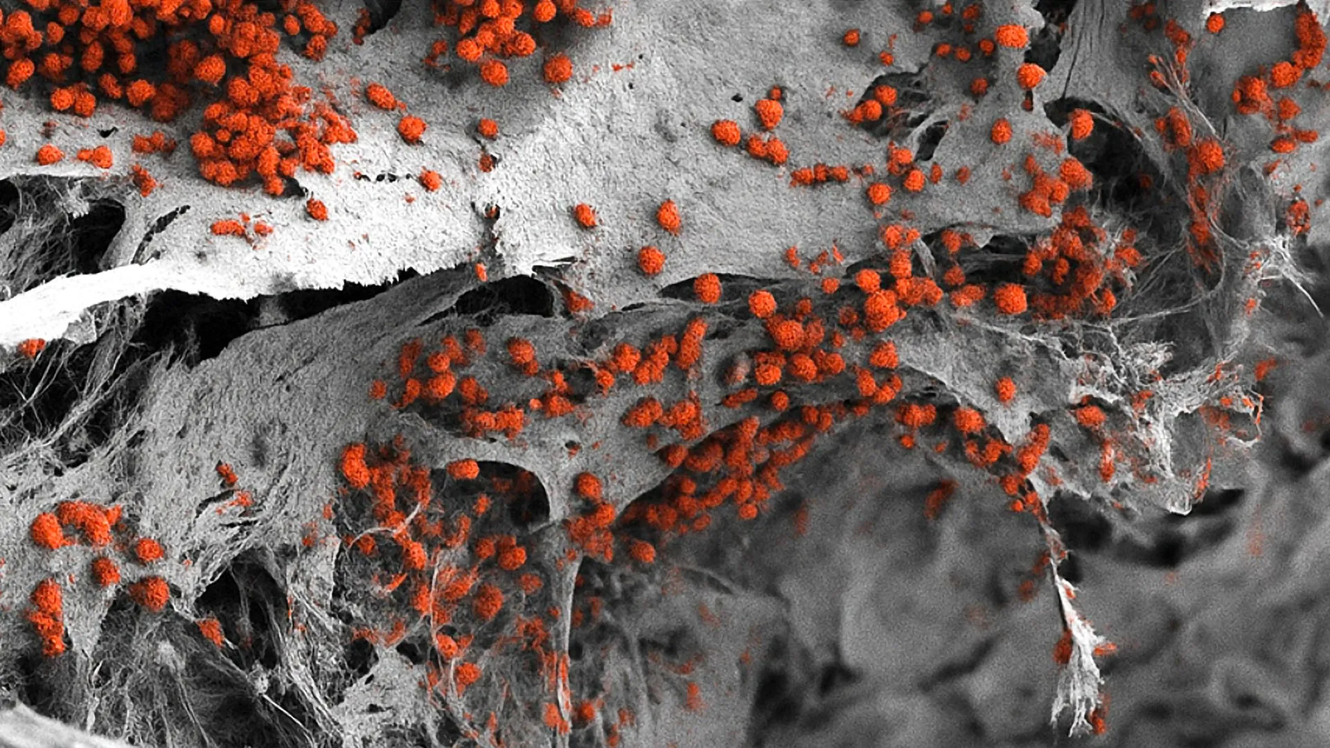
Stem cells are the foundational building blocks of the human body, serving as immature precursors with the unique ability to differentiate into specialized tissue. In the context of the hematological system, blood stem cells—formally known as hematopoietic stem cells (HSCs)—reside primarily within the bone marrow. Under normal physiological conditions, these cells exist in a state of "quiescence." This is a form of cellular hibernation where the cells divide very slowly, a protective mechanism that prevents the exhaustion of the stem cell pool over a person’s lifetime and minimizes the risk of DNA mutations that could lead to cancer.
However, when the body sustains an injury or undergoes significant blood loss, these quiescent cells must rapidly transition into an activated state. During activation, they multiply at high speeds and transform into mature, functional cells, such as oxygen-carrying red blood cells, infection-fighting white blood cells, and clot-forming platelets. The Weill Cornell Medicine study identifies FLI-1 as the critical DNA transcription-regulating protein that governs this transition. Without FLI-1, stem cells remain locked in their dormant state, unable to respond to the body’s regenerative demands.
Identifying FLI-1: The Master Regulator
The research team, led by Dr. Shahin Rafii, director of the Hartman Institute for Therapeutic Organ Regeneration and the Ansary Stem Cell Institute, utilized advanced single-cell profiling and computational analysis to compare the genetic landscapes of quiescent and activated blood stem cells. Their investigation zeroed in on FLI-1, a transcription factor known to control the activity of thousands of downstream genes.
The study revealed that FLI-1’s primary function is to facilitate communication between blood stem cells and their surrounding environment, specifically the "vascular niche." This niche consists of specialized endothelial cells that line the blood vessels within the bone marrow. The researchers demonstrated that when FLI-1 is absent, blood stem cells become "loners," losing their ability to interact with the endothelial cells. This lack of interaction keeps the cells in a permanent state of hibernation. Conversely, when FLI-1 is present, it restores the stem cells’ connections and "co-adaptability" with the vascular niche, pushing them into a regenerative state that allows them to expand their numbers and restore the blood supply.
A Breakthrough in Transplant Efficiency
The implications of this discovery for bone marrow transplants are profound. Bone marrow transplants are a cornerstone of treatment for various blood cancers, such as leukemia and lymphoma, as well as certain genetic disorders. The procedure involves replenishing a patient’s immune and blood cell populations using healthy stem cells. However, the success of these transplants often hinges on the quantity and quality of the donor cells.
"The approach we outlined in this study could substantially improve the efficiency of marrow transplants and marrow-cell-targeted gene therapies, especially in cases where the donor has a very limited supply of viable blood stem cells," said Dr. Shahin Rafii, who also serves as the Arthur B. Belfer Professor in Genetic Medicine at Weill Cornell Medicine.
In many clinical scenarios, particularly when using a patient’s own stem cells (autologous transplants), the cells may have been damaged or weakened by previous rounds of chemotherapy or radiation. These "exhausted" cells are difficult to activate and expand in a laboratory setting. By transiently introducing FLI-1 into these cells, doctors could potentially rejuvenate them, ensuring they are primed for successful engraftment once re-infused into the patient.
The Role of mRNA Technology in Stem Cell Therapy
One of the most innovative aspects of the study is the method used to stimulate the stem cells. While FLI-1 is essential for regeneration, its chronic overactivity is associated with the development of certain types of leukemia. To circumvent this risk, the researchers developed a "hit-and-run" approach.
Using a method similar to the technology found in modified mRNA-based vaccines, the team introduced FLI-1 into the stem cells for only a brief period—typically a few days. This transient pulse of FLI-1 was sufficient to "wake" the cells from their dormant state and initiate the expansion process without causing the long-term genetic changes that could lead to malignancy.
Dr. Tomer Itkin, study co-first author and current director of Tel Aviv University’s Neufeld Cardiovascular Research Institute, emphasized the safety of this approach. "The stem cells we prime with FLI-1 modified mRNA in this way wake up from hibernation, expand and functionally and durably engraft in the recipient host, without any evidence of cancer," Dr. Itkin noted. This suggests that the timing of protein expression is just as important as the protein itself in therapeutic applications.
Solving the Umbilical Cord Blood Puzzle
For decades, hematologists have observed that blood stem cells derived from human umbilical cords possess a significantly higher regenerative potential than those harvested from adult bone marrow or peripheral blood. However, the underlying biological reason for this difference remained elusive until now.
The Weill Cornell team addressed this long-standing puzzle by demonstrating that umbilical cord-derived stem cells naturally exhibit higher levels of FLI-1 activity. This elevated activity allows cord blood cells to interact more effectively with the regenerative vascular niche, explaining their superior potency. By understanding this mechanism, researchers may now be able to "upgrade" adult stem cells to match the performance levels of cord blood cells, effectively expanding the pool of high-quality donor material available for patients.
Chronology and Development of the Research
The path to this discovery was paved by years of investigation into the "niche theory" of stem cells. First proposed in the late 1970s, the theory suggested that stem cells do not operate in a vacuum but are heavily influenced by their immediate microenvironment. The Weill Cornell study provides the most detailed molecular map to date of how this interaction is governed at the genetic level.
- Initial Profiling: The team began with single-cell RNA sequencing to identify gene expression differences between dormant and active cells.
- Target Selection: Bioinformatics analysis identified FLI-1 as a central node in the regulatory network of activated cells.
- Validation: Preclinical models were used to observe the effects of FLI-1 deletion, which resulted in a failure of stem cells to respond to injury.
- Therapeutic Testing: The researchers applied modified mRNA technology to deliver transient FLI-1 pulses to adult human stem cells, observing a marked increase in expansion and engraftment success.
Broader Implications for Gene Therapy and Blood Disorders
Beyond transplants, the discovery of the FLI-1 switch has major implications for gene therapy. Disorders such as beta-thalassemia and sickle cell anemia require the harvesting of a patient’s own stem cells, the insertion of a corrective gene in a laboratory, and the subsequent expansion of those cells before they are returned to the patient.
This process is often fraught with difficulty, as stem cells are notoriously fragile once removed from the body. A safe and reliable method for switching these cells into a regenerative state could drastically reduce the time needed for laboratory expansion and increase the survival rate of the edited cells. This could make gene therapies more accessible and reduce the overall cost of treatment by shortening hospital stays and improving long-term patient outcomes.
Future Directions and Clinical Translation
The research team plans to move forward with further preclinical development, focusing on scaling up the modified mRNA delivery method. The ultimate goal is to transition into human clinical trials, where the FLI-1 activation technique can be tested in a controlled medical environment.
"We showed that stem cell activity is not autonomous but also is not fully determined by endothelial cell vascular niche signals—it depends instead on signaling and adaptability between the two," said co-first author Sean Houghton, a bioinformatics analyst. This nuanced understanding of the "crosstalk" between cells is expected to inform the next generation of regenerative therapies.
As the medical community moves toward precision medicine, the ability to control the "on/off" switch of cellular regeneration offers a powerful tool. If the results of this preclinical study can be replicated in humans, it may set the stage for a new era in hematology, where blood production can be safely and durably restored for a wide range of life-threatening conditions.
The study received support from several branches of the National Institutes of Health (NIH), including the National Heart, Lung, and Blood Institute and the National Institute of Diabetes and Digestive and Kidney Diseases. Additional funding was provided by the Hartman Institute for Therapeutic Organ Regeneration and the Ansary Stem Cell Institute, underscoring the collaborative and multi-institutional nature of this scientific milestone.


Leave a Reply