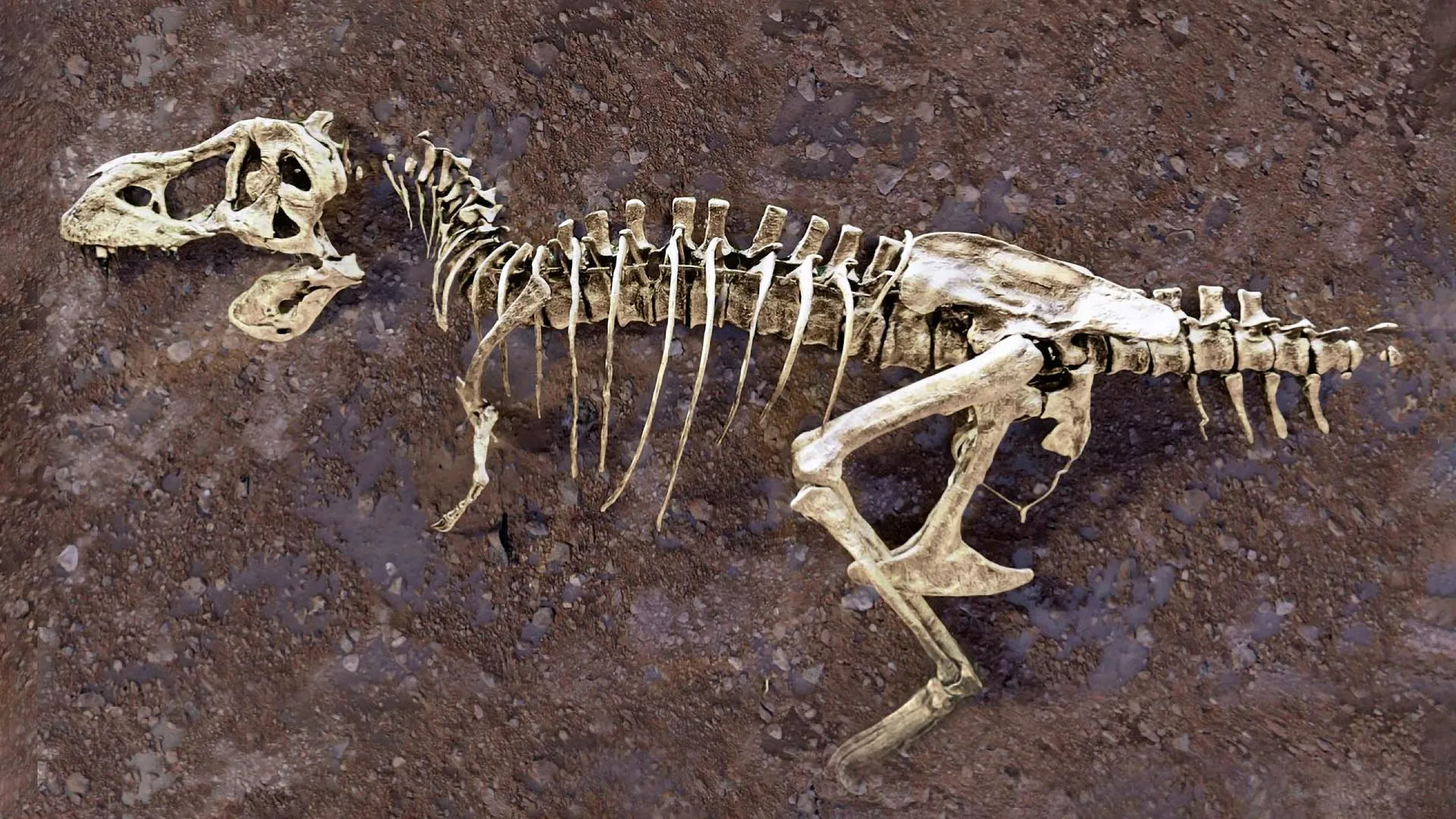
The discovery of "Scotty," the most massive Tyrannosaurus rex skeleton ever unearthed, has long been a centerpiece of paleontological research. However, a recent breakthrough involving a single, unassuming rib bone has elevated the specimen from a marvel of sheer size to a profound biological time capsule. By leveraging advanced neutron imaging at the Department of Energy’s (DOE) Oak Ridge National Laboratory (ORNL), an international team of researchers has successfully peered inside this 66-million-year-old fossil, uncovering a network of mineralized blood vessels that were actively healing at the time of the animal’s death. This discovery provides an unprecedented, high-resolution "medical record" of an apex predator in the final moments of its life during the Late Cretaceous period.
The Anatomy of an Ancient Injury
The rib in question reveals a traumatic fracture that occurred during the final stages of the dinosaur’s life. Typically, soft tissues such as blood vessels, nerves, and organs decompose rapidly following an animal’s death, leaving behind only the mineralized skeletal structure. In this rare instance, the unique environmental conditions of a salty marsh—the site where Scotty eventually perished—served to inhibit the decomposition process, allowing for the partial mineralization of the vascular network.
As the bone fractured, the body’s natural inflammatory response triggered the migration of iron-rich blood to the site of the trauma. New blood vessels began to sprout as part of the regenerative process, a biological mechanism common to vertebrate healing. Because Scotty died before the bone could fully knit back together, the nascent vascular growth was trapped within the fossilized matrix. For modern researchers, this provides a rare snapshot of metabolic activity in a prehistoric beast, offering clues about how these massive theropods managed pain and systemic healing.
A Chronology of Discovery
The path to this discovery began long before the neutron beams of 2026. The specimen itself, Scotty, was first identified by paleontologists from the Royal Saskatchewan Museum in the rugged Frenchman River Valley of Canada. Since its initial excavation, the specimen has been subject to rigorous analysis, but the internal secrets of its ribs remained hidden until 2020.
- 2020: Jerit Mitchell, then an undergraduate student at the University of Regina (U of R), performed the first micro-CT scans of the rib sections at the Canadian Light Source. These X-ray images provided the initial evidence of anomalous internal structures, suggesting the presence of fossilized soft tissue.
- 2021–2023: Researchers expanded their methodology, employing synchrotron radiation and advanced microscopy to map the vascular structures at a cellular level.
- April 2026: The research reached a milestone at the Oak Ridge National Laboratory. The team utilized the Multimodal Advanced Radiography Station (MARS) and the Virtual Environment for Neutron Sciences (VENUS) to non-destructively probe the specimen.
- Present Day: The data obtained through these neutron imaging instruments is currently being synthesized to create a comprehensive model of dinosaurian injury response, with plans to compare these findings against other specimens in global collections.
Technological Synergy: Neutrons vs. X-Rays
The success of this investigation relies on the complementary nature of modern imaging physics. X-rays, which utilize high-energy electromagnetic radiation, are highly effective at visualizing dense, mineralized structures. However, they often struggle to distinguish between subtle variations in soft tissue preservation.
Neutron imaging, by contrast, operates on different physical principles. Neutrons are particularly sensitive to light elements, most notably hydrogen. Because organic matter and residual moisture are rich in hydrogen, neutron beams can highlight structures that are essentially "invisible" to standard X-ray systems. By integrating these two modalities, the team at U of R and ORNL was able to construct a 3D internal map of the rib that differentiates between the original bone mineral and the mineralized remains of the blood vessels.
According to Dr. Mauricio Barbi, a professor of physics at the University of Regina, the quality of the data is unparalleled. "It is like winning the lottery," Barbi noted. The ability to visualize these vessels without destroying the fossil allows the team to preserve the specimen for future generations while extracting data that was previously considered unattainable.
Institutional Collaboration and Scientific Significance
The project represents a high-level collaboration between academic institutions and national laboratories. The University of Regina’s physics department provided the theoretical framework and lead researchers, while the ORNL facilities provided the necessary hardware. Hassina Bilheux, the lead instrument scientist for VENUS, emphasized that the application of neutron science to paleontology is an emerging field with massive potential.
"People often think of neutrons as tools for studying batteries or advanced materials," Bilheux explained. "But they are just as innovative for answering fundamental questions about ancient life." This cross-disciplinary approach is shifting how paleontologists view museum collections. Rather than treating fossils as static objects to be observed, researchers are beginning to treat them as complex, three-dimensional datasets that can be "queried" using high-energy physics.
Broader Implications for Paleontology
The implications of the Scotty rib study extend far beyond a single dinosaur. By confirming that soft tissue structures can be preserved and imaged, the study provides a roadmap for analyzing other specimens held in storage worldwide. Many fossils that were previously discarded or deemed "poorly preserved" due to internal fractures or discoloration may now be subject to re-examination.
Furthermore, the study of injury and healing in dinosaurs allows researchers to infer behavioral patterns. If a large T. rex survived a significant rib fracture, it implies that the animal had the metabolic capacity to endure the pain and the social or ecological support to continue feeding while incapacitated. This challenges earlier assumptions that large theropods were strictly solitary and that significant injuries were invariably fatal.
Marcella Berg, an assistant professor at U of R, highlighted the broader goal of this research: "By piecing the clues together, we understand the past and how things could evolve in the future." As the team continues to process the data from the VENUS and MARS instruments, they are developing a comparative database of pathologies. This database will allow scientists to categorize how different dinosaur species responded to trauma, potentially revealing trends in survival strategies and evolutionary resilience.
Future Directions
The team plans to extend their research to include other materials from the Frenchman River Valley, such as fossilized amber and dinosaur scales. These samples, also rich in hydrogen, are ideal candidates for neutron imaging. The long-term objective is to move beyond the study of a single bone and toward a more holistic understanding of the internal physiology of the Late Cretaceous fauna.
As Jerit Mitchell, the project’s lead doctoral candidate, concluded, there are countless secrets waiting to be uncovered in existing museum collections. The shift toward non-invasive, high-resolution imaging technology ensures that these discoveries will not come at the cost of the integrity of the fossils. As the technology becomes more accessible, the study of dinosaur biology is poised to move from the realm of descriptive anatomy into the realm of high-precision forensic science, fundamentally changing our understanding of the life and death of the titans that once ruled the Earth.