The complex architecture of the bone marrow, long considered one of the most challenging biological environments to visualize in its native state, has been successfully mapped with unprecedented detail by a team of researchers at the Indiana University School of Medicine. By leveraging a sophisticated multiplex imaging platform, the scientists have developed a methodology to observe dozens of cellular markers simultaneously within intact mouse bone marrow tissue. This breakthrough, recently detailed in the journal Leukemia, addresses a decades-old limitation in hematological research and opens new avenues for the study of blood cancers, immune disorders, and regenerative medicine.
The Challenge of the Bone Marrow Microenvironment
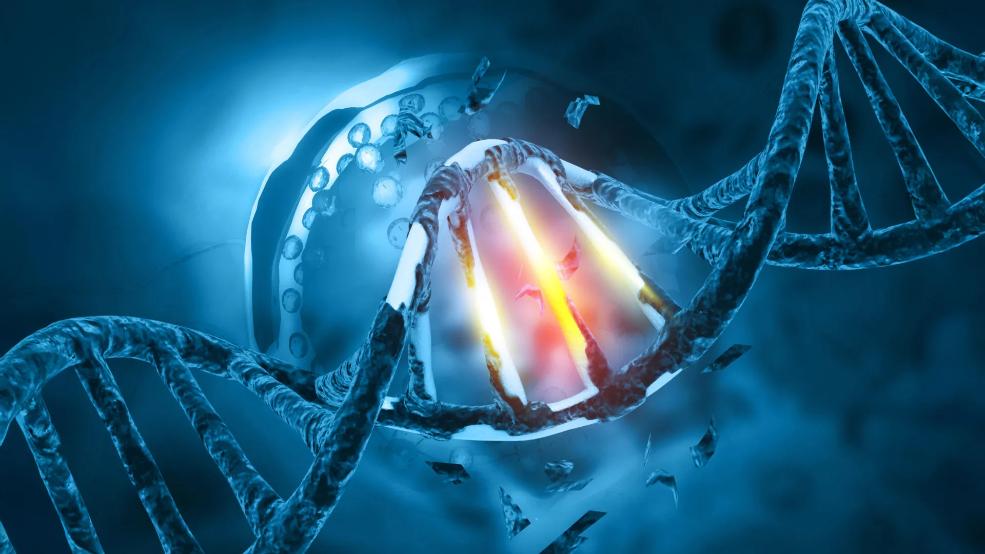
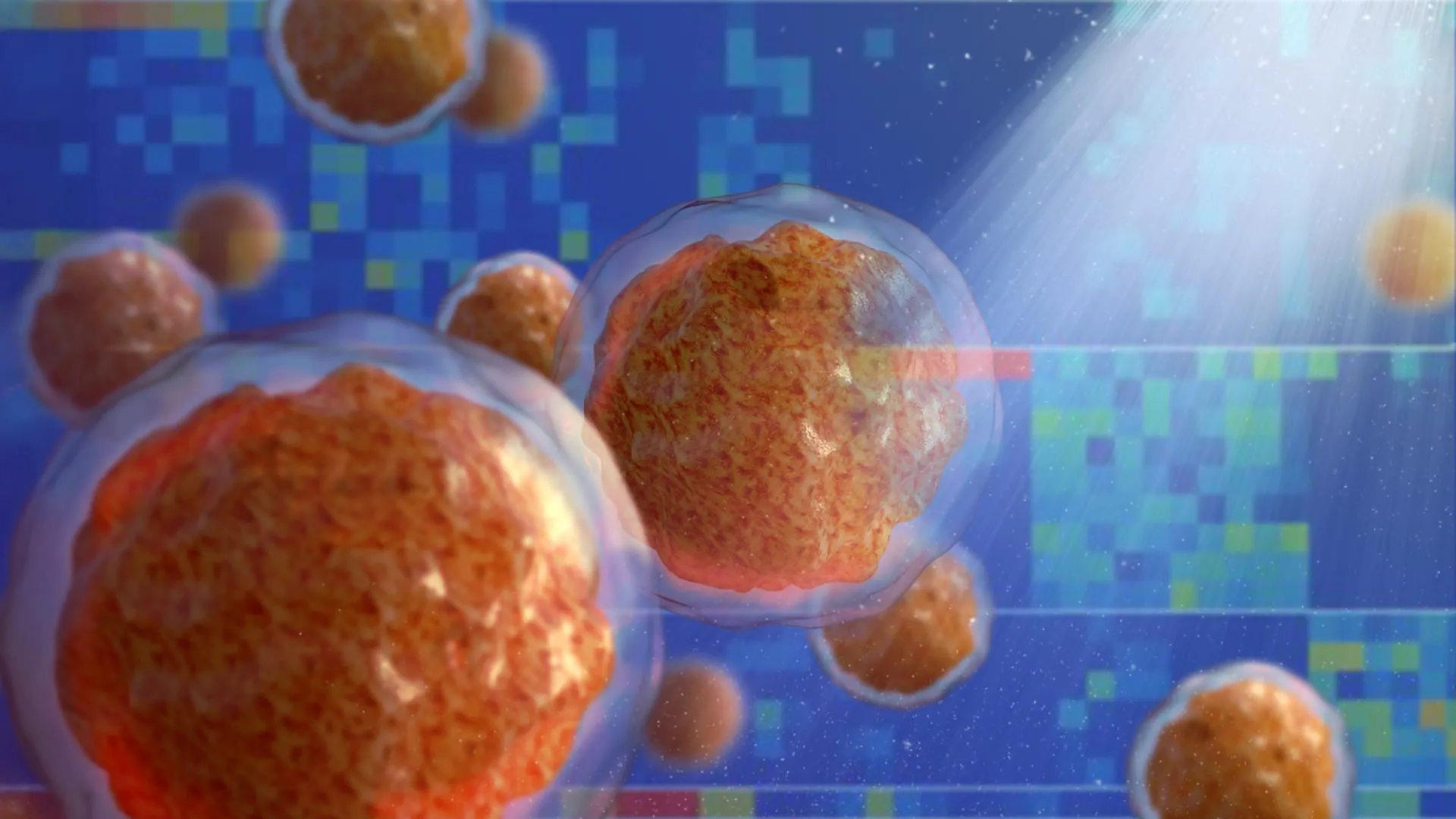
Bone marrow is the primary site of hematopoiesis—the process by which the body produces new blood cells. It serves as a sanctuary for hematopoietic stem cells (HSCs), which differentiate into oxygen-carrying red blood cells, infection-fighting white blood cells, and clot-forming platelets. However, studying this process in its natural setting has historically been hindered by the physical properties of the tissue itself.
The marrow is a soft, gelatinous substance sequestered within the rigid, calcified walls of the cortical bone. This "black box" nature of the skeletal system has forced researchers to choose between two suboptimal options: destroying the tissue structure to count its components or observing a very limited number of components within the preserved structure.
Sonali Karnik, PhD, an assistant research professor of orthopedic surgery at the IU School of Medicine and co-lead author of the study, emphasized the significance of overcoming these barriers. "Bone marrow is difficult to study because it is gelatinous and encased in hard bone," Dr. Karnik stated. "Since bone marrow plays an important role in blood and immune cell formation and houses valuable stem cells, our unique imaging approach offers a useful tool for a variety of research applications."
Technological Evolution: From Triple-Color to Multiplex Imaging
For decades, the gold standard for analyzing bone marrow has been flow cytometry. While highly effective at quantifying specific cell populations, flow cytometry requires the bone to be crushed and the marrow to be processed into a liquid suspension. This "smoothie" approach provides a census of the cells present but completely erases the "geography" of the tissue. Researchers lose critical information regarding which cells are neighbors, how they are positioned relative to blood vessels, and how the surrounding bone matrix influences their behavior.
Standard fluorescence microscopy, the alternative, allows for the preservation of tissue architecture. However, it is traditionally limited by the "spectral overlap" of fluorescent dyes, meaning scientists can typically only visualize three to four different cellular markers at one time. In a tissue as complex as bone marrow, where dozens of cell types—including osteoblasts, endothelial cells, adipocytes, and various stages of maturing immune cells—interact, a four-color limit provides only a keyhole view of a vast landscape.
The IU research team utilized the Phenocycler 2.0 (formerly known as CODEX), a multiplex imaging tool that utilizes DNA-barcoded antibodies. This technology allows for iterative cycles of staining and imaging, enabling the researchers to visualize a record-breaking 25 different cellular markers in a single, intact tissue section. By maintaining the spatial integrity of the marrow, the team can now see not just what cells are there, but exactly where they are located and how they are interacting with their microenvironment, or "niche."
Chronology of Development and Experimental Milestones
The development of this imaging protocol was a multi-year effort spearheaded by the IU Cooperative Center of Excellence in Hematology (CCEH). The project followed a rigorous timeline of optimization to adapt technology previously used for softer organs to the unique demands of bone-encased tissue.
- Initial Adaptation (2021-2022): The team began by testing the Phenocycler platform on soft tissues like the spleen and kidney. These organs lack the calcified barriers of bone, making them ideal for establishing the baseline parameters for the DNA-barcoded antibody panels.
- Optimizing Bone Sectioning (2022-2023): The primary hurdle was creating ultra-thin, high-quality sections of mouse bone that retained the delicate marrow inside. Traditional decalcification processes often degrade cellular proteins, making them invisible to antibodies. The IU team refined a specialized sectioning and fixation technique that preserved both the bone mineral and the delicate cellular antigens.
- Marker Panel Validation (Late 2023): The researchers curated a panel of 25 antibodies specifically designed to identify various lineages of the hematopoietic system and the structural cells of the bone marrow niche. Each antibody had to be validated to ensure it would bind correctly under the multiplexing conditions.
- Publication and Patent Filing (2024): Following the successful imaging of the mouse marrow, the findings were submitted to Leukemia. Simultaneously, the IU Innovation and Commercialization Office filed a provisional patent for the specific methodology used to prepare and image the bone marrow, recognizing its potential as a commercial and diagnostic tool.
Supporting Data and Technical Breakthroughs
The data generated by the Phenocycler 2.0 system provides a high-dimensional map of the marrow. In their published study, the IU researchers demonstrated the ability to identify and localize rare stem cell populations that make up less than 0.01% of the total marrow volume.
Key data points from the study include:
- Marker Density: The visualization of 25 markers simultaneously represents an eightfold increase in data density compared to standard four-color immunofluorescence.
- Spatial Resolution: The technique maintains sub-cellular resolution, allowing researchers to observe the proximity of leukemia cells to specific "protective" cells in the niche that may contribute to drug resistance.
- Tissue Integrity: Unlike flow cytometry, which has a 0% retention of spatial data, the new method provides 100% spatial mapping of the analyzed area.
This high-dimensional data allows for "spatial proteomics," where researchers can use computational algorithms to identify "neighborhoods" within the bone marrow. For example, they can determine if certain immune cells preferentially cluster near blood vessels (perivascular niches) or near the bone surface (endosteal niches) during different stages of disease.
Official Responses and Collaborative Impact
The success of the project is attributed to the collaborative environment at the IU School of Medicine, particularly the synergy between the Department of Orthopedic Surgery and the Herman B Wells Center for Pediatric Research.
Reuben Kapur, PhD, a co-senior author on the study and director of the Herman B Wells Center, highlighted the broader implications for human health. "Because mouse models are widely used to study human diseases, this technique offers a promising new method for investigating a range of conditions like autoimmune diseases, leukemia and other disorders involving bone marrow," Kapur said.
As the co-director of the IU Cooperative Center of Excellence in Hematology, Kapur noted that the center is the first in the world to successfully apply this specific multiplexing tool to mouse bone marrow. This achievement positions Indiana University as a global leader in spatial hematology.
The research also involved a diverse team of specialists, including experts in pediatric oncology, regenerative medicine, and bioinformatics. The interdisciplinary nature of the team—including authors like Melissa A. Kacena and Karen E. Pollok—underscores the multifaceted utility of the imaging technique, from understanding how bones heal to how pediatric cancers metastasize.
Broader Impact: From Cancer Research to Drug Development
The implications of this imaging breakthrough extend far beyond basic laboratory science. In the realm of oncology, particularly leukemia research, the "niche" is a critical factor. It is well-documented that cancer cells can "hijack" the bone marrow microenvironment, using normal cells to protect themselves from chemotherapy. By using the IU imaging technique, drug developers can now see exactly how a new therapy disrupts these protective neighborhoods, potentially leading to more effective treatments that flush cancer cells out of their hiding spots.
In autoimmune research, the technique could be used to study how "memory" B cells persist in the bone marrow for decades, sometimes driving chronic inflammation. Understanding the spatial cues that allow these cells to survive could lead to breakthroughs in treating lupus, rheumatoid arthritis, and multiple sclerosis.
Furthermore, the technique has significant applications for musculoskeletal disorders. By expanding the marker panel to include bone, nerves, and muscle, as the team is currently doing, researchers can study how the "neuro-vascular-bone" unit functions as a whole. This is particularly relevant for aging populations suffering from osteoporosis or for patients recovering from traumatic bone injuries.
Future Directions and Scaling the Technology
The IU School of Medicine team is not stopping at 25 markers. Current efforts are focused on expanding the panel to include more signaling molecules and structural components. The goal is to create a "holistic map" that includes the nervous system’s interaction with the bone marrow, a field known as neuro-immunology.
"The team is now working to expand the marker panel to include additional features such as bone, nerves, muscle and more immune and signaling cell types," the researchers noted. This expansion will likely involve integrating the protein-level data from the Phenocycler with spatial transcriptomics, which measures gene activity.
While the current study utilized mouse models, the logical next step is the translation of this protocol to human clinical samples. High-resolution spatial mapping of human bone marrow biopsies could eventually become a diagnostic standard, allowing pathologists to see the "spatial signatures" of disease long before they become apparent through traditional blood tests.
Supported by funding from the National Institutes of Health (NIH), this research represents a fundamental shift in how scientists interact with the skeletal system. By turning the "black box" of bone marrow into a transparent, high-definition map, the IU School of Medicine has provided the global scientific community with a powerful new lens through which to view the very foundations of human health and disease.