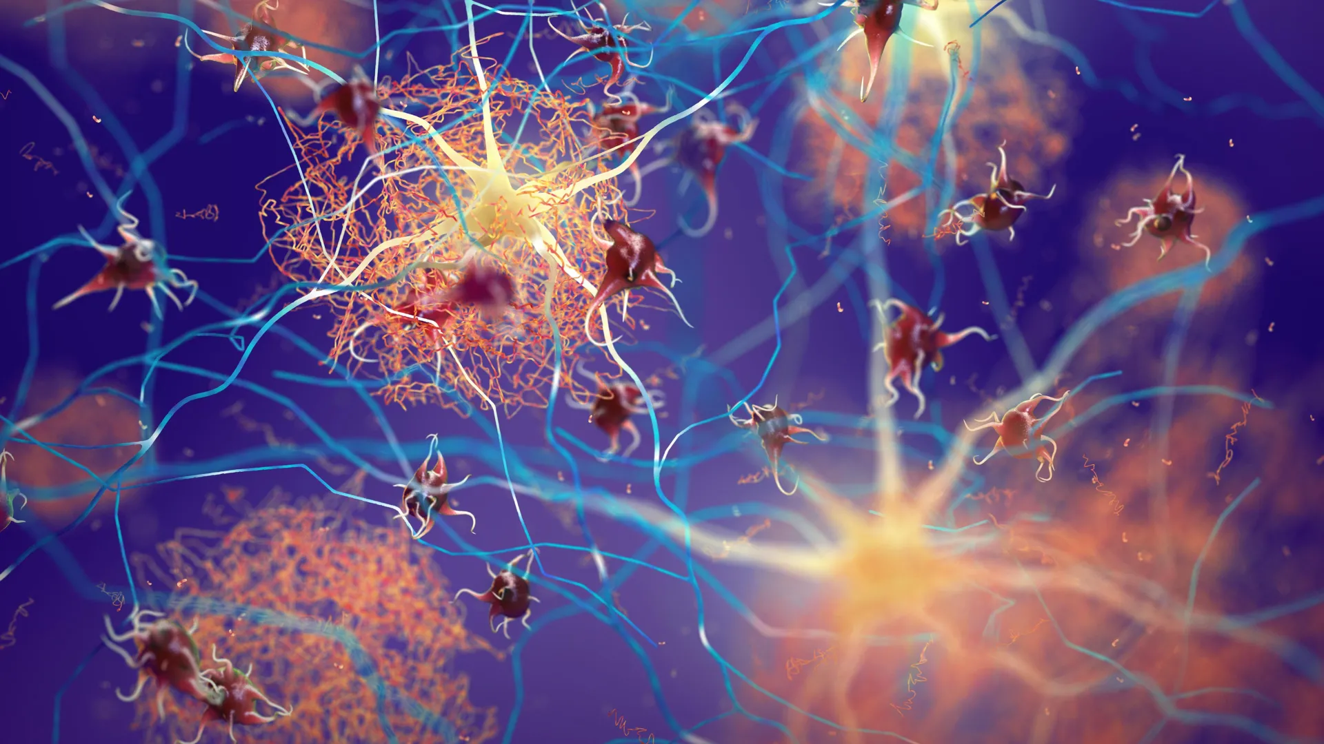
The intricate architecture of the human brain begins as a bustling construction site where billions of newly formed neurons must navigate a labyrinthine environment to reach their designated positions. During this developmental phase, these cells are forced to squeeze through microscopic gaps in densely packed tissue, a process that researchers have long viewed as a purely physical feat of navigation. However, a groundbreaking study published in the journal Nature has revealed that this journey comes at a significant biological cost: the physical strain of migration causes extensive damage to the neurons’ DNA. This discovery, led by the Institute for Integrated Cell-Material Sciences (WPI-iCeMS) at Kyoto University, suggests that DNA breakage is not merely a byproduct of cellular stress but a routine occurrence in healthy brain development.
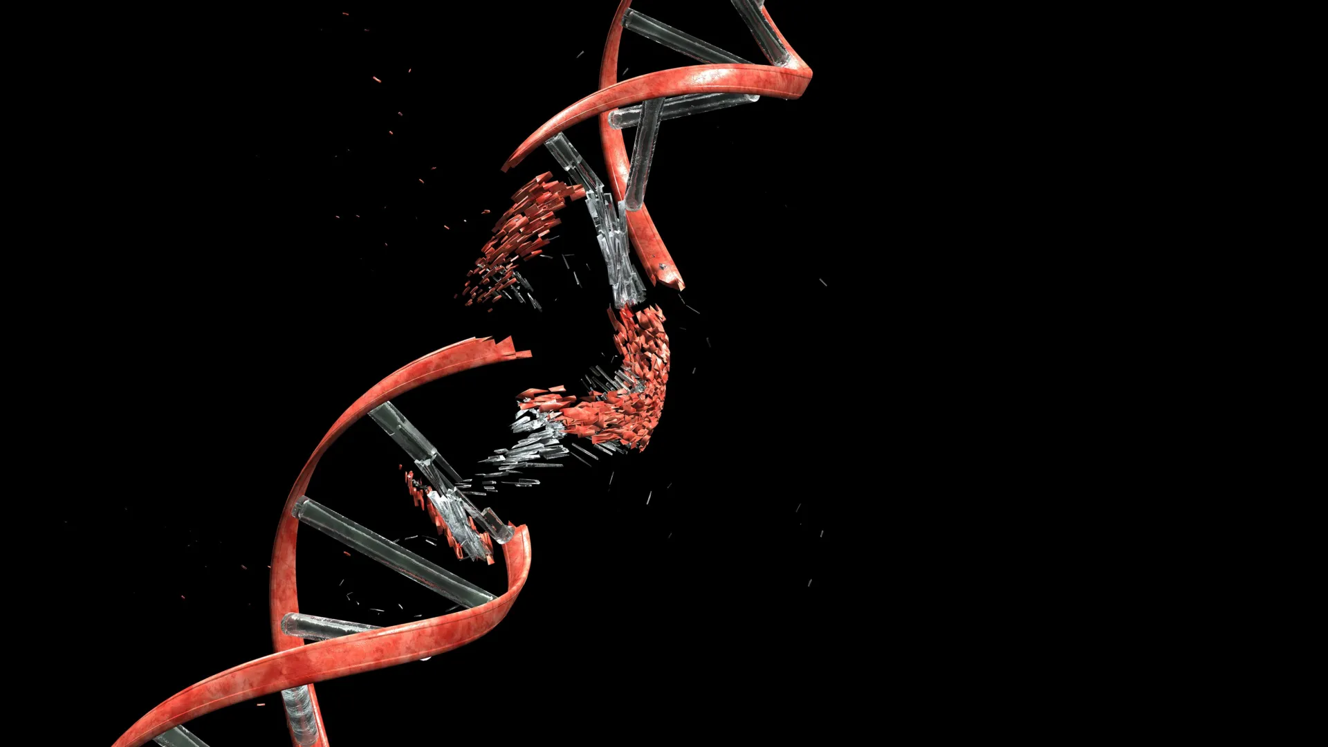
The research team, which included collaborators from the University of Tokyo, Osaka University, the National University of Singapore, and the Tokyo Metropolitan Institute of Medical Science, identified that as neurons migrate toward the cerebral cortex, they frequently experience double-strand breaks (DSBs). DSBs are among the most severe forms of genomic instability, occurring when both strands of the DNA double helix are severed. While such damage is typically associated with aging, cancer, or cell death, this study demonstrates that the developing brain possesses highly efficient mechanisms to repair these breaks, ensuring that the neurons remain functional despite the structural trauma of their journey.
The Physical Challenge of Brain Assembly
The cerebral cortex is the seat of higher cognitive functions, including thought, memory, and voluntary movement. For this structure to form correctly, neurons generated in the deeper layers of the developing brain must travel outward along radial glial fibers. This migration is a high-stakes obstacle course. The cells must push through a matrix of existing fibers, extracellular material, and other neighboring cells.
To understand the impact of this physical environment on the integrity of the neuronal genome, the research team developed a sophisticated experimental model. They utilized microfluidic technology to create microchannels that mimicked the narrow, confined spaces of the developing embryonic brain. By guiding living neurons through these channels, which were significantly narrower than the cells themselves, the scientists could observe the cellular response to mechanical compression in real-time.
Using advanced fluorescent markers designed to light up in the presence of DNA damage signals, the team observed a striking phenomenon. As the nucleus of the neuron—the compartment housing the cell’s genetic blueprint—was squeezed through the narrowest points of the microchannels, markers for double-strand breaks spiked. The physical deformation of the nucleus appeared to be the primary driver of this genomic instability.
The Role of Topoisomerase IIβ as a Molecular Culprit
A central finding of the study involves the enzyme Topoisomerase IIβ (Top2β). In normal cellular life, Top2β is essential for managing the topological state of DNA. As the double helix unwinds for replication or gene expression, it can become over-wound or tangled, much like a telephone cord. Top2β acts as molecular scissors, cutting the DNA to relieve this tension and then immediately rejoining the strands.
The researchers discovered that the mechanical stress of migration disrupts this delicate "cut-and-paste" cycle. When a neuron is squeezed, the physical pressure appears to trap Top2β in the middle of its task. Instead of completing the repair, the enzyme remains bonded to the DNA at the site of the cut, leaving the double-strand break open.
"The process can be compared to cutting a tangled cable to remove twists and then being interrupted before you can reconnect it," explained Professor Mineko Kengaku of WPI-iCeMS, the study’s lead author. "In the context of the migrating neuron, the mechanical journey itself acts as the interruption, leaving the DNA in a fractured state."
A Tale of Two Cells: Neurons versus Cancer
One of the most significant aspects of the research is the comparison between how neurons and cancer cells handle migration-induced DNA damage. Previous studies have shown that when cancer cells metastasize and squeeze through narrow gaps in the body, they also experience DNA damage. However, in cancer cells, this damage is often catastrophic and chaotic. It frequently leads to mutations that can make the cancer more aggressive or trigger apoptosis (programmed cell death).
The Kyoto University team found that neurons are far more resilient. Unlike the random damage seen in cancer, the DNA breaks in migrating neurons were found to be concentrated in specific, non-essential regions of the genome. These "safe zones" are typically areas that do not contain critical instructions for cellular survival or basic function.
Furthermore, the study highlighted the efficiency of the neuronal repair system. Once the neurons emerged from the microchannels, the fluorescent markers for DNA damage began to fade rapidly. The researchers found that the vast majority of the double-strand breaks were successfully repaired within 24 hours. This repair is primarily handled by a pathway known as non-homologous end joining (NHEJ), which acts as a rapid-response team to stitch broken DNA ends back together.
Consequences of Repair Failure: Insights from Mouse Models
To investigate what happens when this natural repair process fails, the researchers conducted experiments using genetically modified mice. These mice were engineered to lack Ligase 4, an enzyme that is a crucial component of the NHEJ repair pathway, specifically within their newly formed cerebellar neurons.
Initially, the mice appeared to develop normally. They reached adulthood without any obvious physical deformities or cognitive deficits. However, as the mice aged, the researchers observed the onset of mild but progressive neurological symptoms. Specifically, the mice began to exhibit balance and coordination issues, symptoms that are characteristic of ataxia—a group of disorders that affect the cerebellum.
This finding is particularly relevant to human medicine. There are several known human genetic disorders, such as Ataxia-telangiectasia, which are caused by deficiencies in DNA repair mechanisms. Patients with these conditions often experience progressive neurological decline. The study provides a potential explanation for why these symptoms often appear later in life: the neurons may survive the initial journey with "unrepaired" or "misrepaired" DNA, but the accumulated genomic instability eventually leads to cellular dysfunction and the breakdown of neural circuits.
Philosophical and Scientific Implications: The Genomic Record of Migration
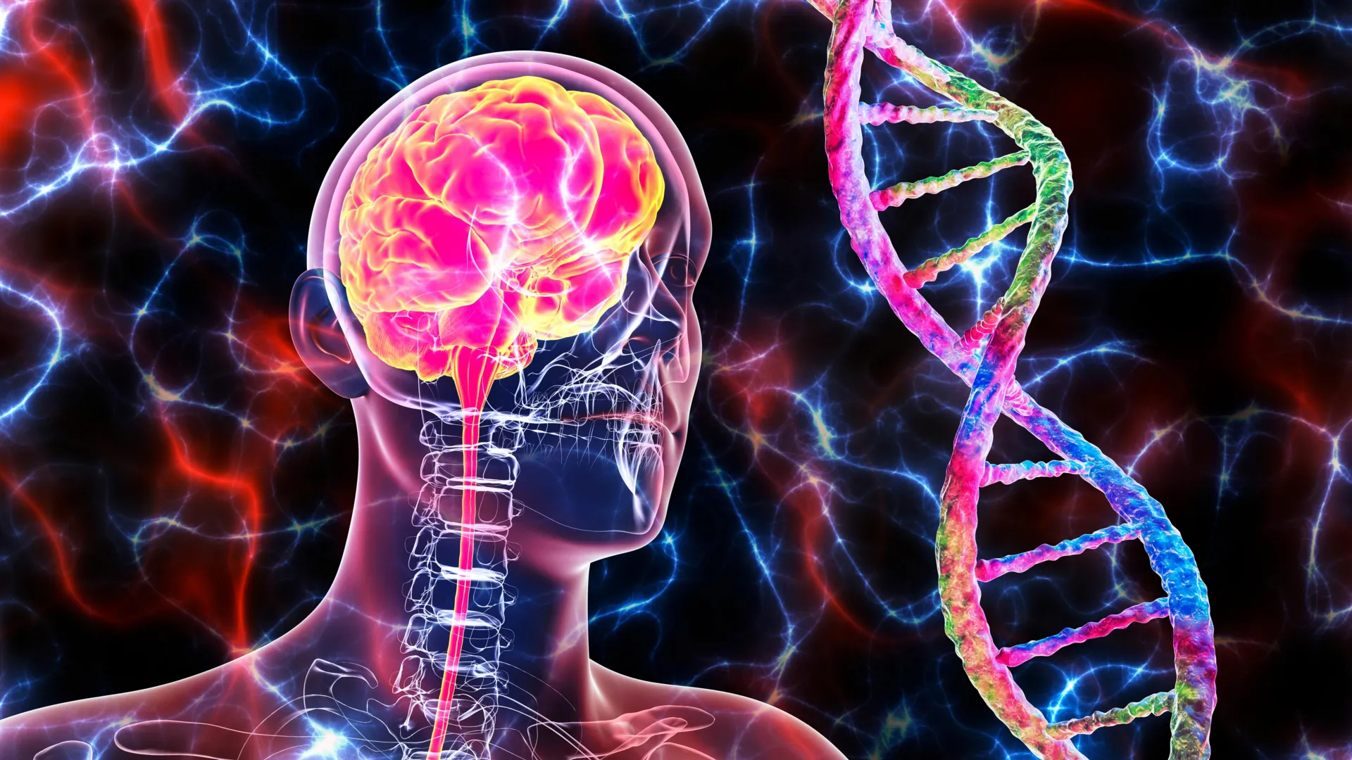
The discovery that DNA damage is a routine part of brain development shifts the paradigm of how scientists view the neuronal genome. Historically, it was assumed that every neuron in an individual’s brain shared the exact same genetic code. However, this study suggests that the physical history of a neuron—the specific path it took and the obstacles it encountered—might leave a permanent mark on its DNA.
"All neurons originate from the same DNA, but DNA damage and repair can introduce small genetic differences between individual neurons through a small mechanical journey," said Professor Kengaku. "Some of that history may be written into the genome itself."
This concept, known as somatic mosaicism, suggests that the brain is a mosaic of cells with slightly different genetic profiles. While the study found that neurons generally repair their DNA successfully, the repair process is not always perfect. Small variations or "scars" left behind by the NHEJ pathway could contribute to the vast diversity of neuronal types and functions found in the adult brain.
Future Research and Clinical Potential
The implications of this research extend far beyond basic developmental biology. By identifying the specific mechanisms by which neurons protect their DNA, scientists may be able to develop new strategies for treating neurodevelopmental and neurodegenerative diseases.
Future research will likely focus on several key areas:
- Thresholds of Tolerance: Determining the exact point at which mechanical stress exceeds a neuron’s ability to repair itself. This could be crucial in understanding how prenatal stressors, such as infections or malnutrition, might impact brain development.
- Long-term Effects of Somatic Mutations: Investigating whether the "scars" left by DNA repair in early development contribute to susceptibility to diseases like Alzheimer’s or Parkinson’s later in life.
- Therapeutic Targets: Exploring whether enhancing the NHEJ pathway or stabilizing Top2β could prevent damage in individuals with known genetic predispositions to DNA repair deficiencies.
The collaborative effort behind this study underscores the complexity of the brain. By combining expertise in cell biology, material science, and genetics, the team has illuminated a hidden chapter of the brain’s origin story. It appears that the very struggle of becoming a part of the brain’s network—the "mechanical journey" through the crowded cellular landscape—is a defining moment that shapes the genetic identity of every neuron.
As neuroscience moves forward, this study serves as a reminder that the physical environment of a cell is just as important as its chemical environment. The journey to the cerebral cortex is not just a change in location; it is a transformative event that tests the limits of cellular endurance and leaves a lasting legacy on the most complex organ in the known universe.