
The human bone marrow is often described as the body’s "blood factory," a highly sophisticated and dynamic tissue responsible for the continuous production of billions of new blood cells every day. This biological engine is not merely a collection of cells but a complex architectural network composed of specialized bone cells, intricate nervous systems, a dense web of blood vessels, and various supportive cell types. For decades, the sheer complexity of this environment has made it nearly impossible to replicate outside the human body. However, a groundbreaking study from the University of Basel and University Hospital Basel has changed this trajectory. Researchers have successfully engineered a functional, three-dimensional bone marrow model using exclusively human cells, marking a significant milestone in regenerative medicine and oncology.
This new laboratory-grown system, detailed in the journal Cell Stem Cell, offers a high-fidelity replica of the human bone marrow environment. Led by Professor Ivan Martin and Dr. Andrés García-García from the Department of Biomedicine, the research team has provided the scientific community with a tool that could revolutionize how we study blood-related diseases, test new pharmaceuticals, and potentially develop personalized treatments for patients suffering from leukemia and other hematological malignancies.
The Biological Context: Understanding the Bone Marrow Niches
To appreciate the magnitude of this achievement, one must understand the vital role bone marrow plays in human health. Nestled within the cavities of large bones, the marrow is the primary site of hematopoiesis—the process of blood cell formation. Within this environment, hematopoietic stem cells (HSCs) differentiate into red blood cells, white blood cells, and platelets.
However, this process does not happen in a vacuum. It occurs within specific microenvironments known as "niches." These niches provide the physical scaffolding and chemical signals necessary for stem cells to survive, proliferate, or differentiate. Among these, the "endosteal niche" is of particular interest to researchers. Located near the inner surface of the bone, the endosteal niche is a crossroads where bone cells (osteoblasts), blood vessels (endothelial cells), and nerve fibers interact.
Crucially, the endosteal niche is also believed to be a "sanctuary" for cancer cells. In cases of acute myeloid leukemia or other blood cancers, malignant cells often retreat to this niche, where the unique microenvironment protects them from chemotherapy and other treatments. Understanding why this protection occurs has been a primary goal of oncology, but until now, scientists lacked a human-specific model that included all the necessary components—bone, vessels, and nerves—in a single integrated system.
The Limitations of Traditional Research Models
For over half a century, the study of bone marrow and blood diseases has relied heavily on two primary methods: animal models (predominantly mice) and two-dimensional (2D) cell cultures. While these methods have yielded significant insights, they possess inherent flaws that have slowed the progress of human clinical applications.
Animal models, while complex and systemic, do not always mirror human biology accurately. There are fundamental differences in the genetic expression, immune responses, and signaling pathways between mice and humans. Consequently, many drugs that appear successful in mouse trials fail to show efficacy or safety when they reach human clinical trials. Furthermore, ethical concerns regarding the use of animals in research have led to a global push for the "3Rs"—Replacement, Reduction, and Refinement of animal testing.
On the other hand, 2D cell cultures—where cells are grown in flat plastic dishes—are too simplistic. They lack the three-dimensional architecture, mechanical stiffness, and multi-cellular interactions found in the body. Without these spatial cues, cells often behave differently than they would in a living organism, leading to misleading data. The University of Basel’s 3D model bridges this gap, providing a human-centric environment that maintains the spatial and functional integrity of the tissue.
Methodology: Building a Living System from Scratch
The creation of this model was a multi-stage process that combined material science with advanced stem cell biology. The chronology of the project involved the development of a structural scaffold followed by the biological "seeding" and maturation of the tissue.
The foundation of the model is an artificial bone framework made of hydroxyapatite. Hydroxyapatite is a naturally occurring mineral form of calcium apatite that makes up the bulk of human bone and tooth enamel. By using this material, the researchers provided the cells with a chemically and mechanically familiar environment.
The biological components were derived from human induced pluripotent stem cells (iPSCs). These are adult cells that have been genetically reprogrammed back into an embryonic-like state, giving them the ability to become any cell type in the body. Using specific growth factors and signaling molecules, the researchers guided these stem cells through a series of developmental "checkpoints" to transform them into the various components of the endosteal niche, including mesenchymal cells (which form bone and fat), endothelial cells (which form blood vessels), and neural-like cells.
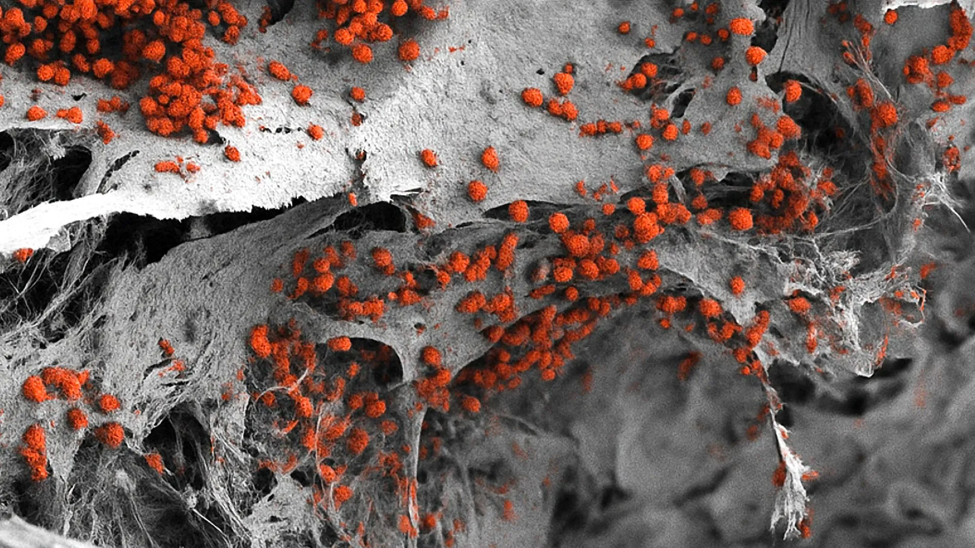
Once introduced into the hydroxyapatite scaffold, these cells organized themselves into a complex 3D structure. Over a period of several weeks, the researchers monitored the development, ensuring that the cells were not just surviving, but actively interacting. The result was a functional piece of human tissue that successfully replicated the signaling and structural characteristics of a natural bone marrow niche.
Technical Specifications and Supporting Data
The Basel model is notable not only for its composition but also for its scale and longevity. While previous attempts at "bone-on-a-chip" technology resulted in microscopic samples, this new model is relatively large, measuring approximately eight millimeters in diameter and four millimeters in thickness. This macro-scale size allows for easier manipulation and observation, making it more practical for laboratory use.
Data published in the study confirmed several key performance indicators:
- Longevity: The system was able to maintain the formation of human blood cells (hematopoiesis) for several weeks. This is a significant improvement over previous models, which often saw cell populations decline rapidly.
- Cellular Diversity: Analysis showed a heterogeneous population of cells, including osteoblasts, pericytes, and endothelial cells, arranged in a way that mimicked the natural architecture of the endosteal niche.
- Biological Fidelity: The researchers observed that the hematopoietic stem cells within the model responded to external stimuli in a manner consistent with human physiology, rather than the "stressed" responses often seen in 2D cultures.
Ethical Progress: Aligning with the 3Rs Principle
One of the most significant implications of this research is its potential to drastically reduce the reliance on animal experimentation. The University of Basel has been a vocal proponent of the 3Rs principle—Replace, Reduce, and Refine.
"We have learned a great deal about how bone marrow works from mouse studies," Professor Ivan Martin noted during the announcement of the findings. "However, our model brings us closer to the biology of the human organism."
By providing a more accurate human-based alternative, the researchers hope to replace many of the preliminary animal tests currently required in hematology and immunology. In the future, researchers could use these synthetic niches to study how the human immune system develops or how viruses interact with bone marrow without ever needing an animal subject. This not only addresses ethical concerns but also increases the scientific validity of the results, as the data is derived from human cells.
Pharmaceutical and Clinical Implications
The pharmaceutical industry stands to benefit immensely from this development. Currently, the "attrition rate" for new drugs—the percentage of drugs that fail during development—is staggeringly high, often exceeding 90%. A primary reason for this failure is that human toxicity or lack of efficacy is only discovered late in the process, during human trials.
A realistic 3D human bone marrow model allows for "high-content screening" early in the drug development cycle. Scientists can introduce experimental compounds into the model and observe how they affect blood cell production or whether they cause damage to the bone marrow niche.
However, Dr. Andrés García-García pointed out a current limitation: "For this specific purpose, the size of our bone marrow model might be too large." To be used for testing hundreds or thousands of drug variations simultaneously (high-throughput screening), the team is now working on miniaturizing the system. A smaller version would allow for automated testing using robotic systems, significantly accelerating the pace of drug discovery.
The Future of Personalized Medicine
Looking further ahead, the most exciting application of this technology lies in personalized medicine. In the current medical landscape, cancer treatment is often a process of trial and error. Two patients with the same type of leukemia might respond very differently to the same chemotherapy regimen.
The researchers envision a future where a "digital twin" or "biological surrogate" of a patient’s bone marrow is created in the lab. By taking a small sample of a patient’s own cells and using them to populate the 3D hydroxyapatite scaffold, doctors could create a patient-specific bone marrow model. They could then test various combinations of drugs on that specific model to see which one most effectively kills the cancer cells while sparing the healthy marrow.
This approach would remove the guesswork from oncology, allowing for the design of "tailor-made" therapies that maximize the chances of remission while minimizing side effects. While the team acknowledges that several technical hurdles remain—such as the time required to grow the tissue and the cost of the process—this study represents the essential first step toward that reality.
Analysis of Broader Impact
The success of the Basel team reflects a broader trend in biotechnology toward "Organ-on-a-Chip" and complex 3D tissue engineering. As we move away from the limitations of the 20th-century research paradigm, the ability to recreate human organ systems in vitro is becoming the new gold standard.
The implications extend beyond cancer. This model could be used to study rare genetic blood disorders, the effects of radiation on the human body, and the aging process of the immune system. It also provides a platform for testing gene therapies, allowing researchers to see how modified stem cells integrate into a realistic human environment before they are ever injected into a patient.
In summary, the creation of a fully human, 3D bone marrow model by the University of Basel and University Hospital Basel is a landmark achievement. By integrating bone, blood vessels, and nerves into a functional system, the researchers have unlocked a new level of biological understanding. While the journey from the laboratory bench to the patient’s bedside is long, the foundation has been laid for a more ethical, accurate, and personalized era of medical science. The "blood factory" is no longer a black box; it is a system that can now be built, observed, and mastered in the laboratory.


Leave a Reply