In a landmark achievement for regenerative medicine and developmental biology, researchers from the Wellcome Sanger Institute and their international collaborators have unveiled the first comprehensive "blueprint" of human skeletal development. This spatial and single-cell atlas, published in the journal Nature on November 20, 2024, provides an unprecedented view of how the human skeleton forms during the earliest stages of life. By mapping the intricate pathways and cellular interactions that occur between five and eleven weeks post-conception, the study offers transformative insights into the origins of common conditions such as osteoarthritis and rare congenital disorders like craniosynostosis. This research is part of the broader Human Cell Atlas (HCA) initiative, a global effort to map every cell type in the human body to better understand health and disease.
The study serves as a critical milestone in a collection of more than 40 HCA publications released simultaneously across Nature Portfolio journals. These papers collectively represent a "leap" in human biological understanding, providing the scientific community with high-resolution data that could redefine how we approach drug safety during pregnancy, joint repair, and the treatment of skeletal deformities.
Mapping the Foundations of the Human Frame
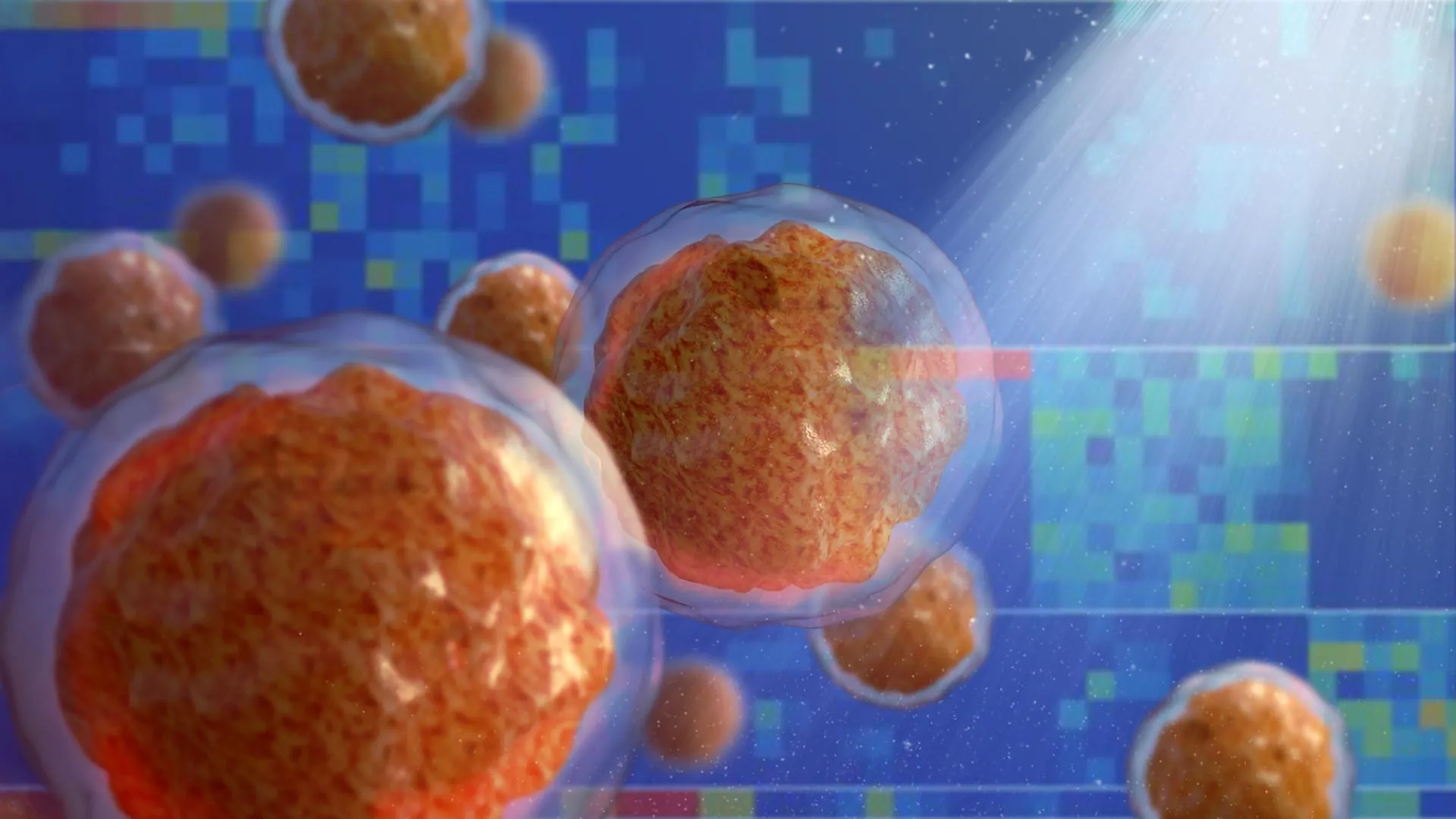
The development of the human skeleton is a highly orchestrated process that begins shortly after conception. Traditionally, our understanding of this process was limited to histological observations—viewing tissues under a microscope to see their shape and structure. However, the new atlas utilizes cutting-edge genomic techniques, including single-cell RNA sequencing and spatial transcriptomics, to identify the specific gene expressions and locations of every cell involved in early bone and cartilage formation.
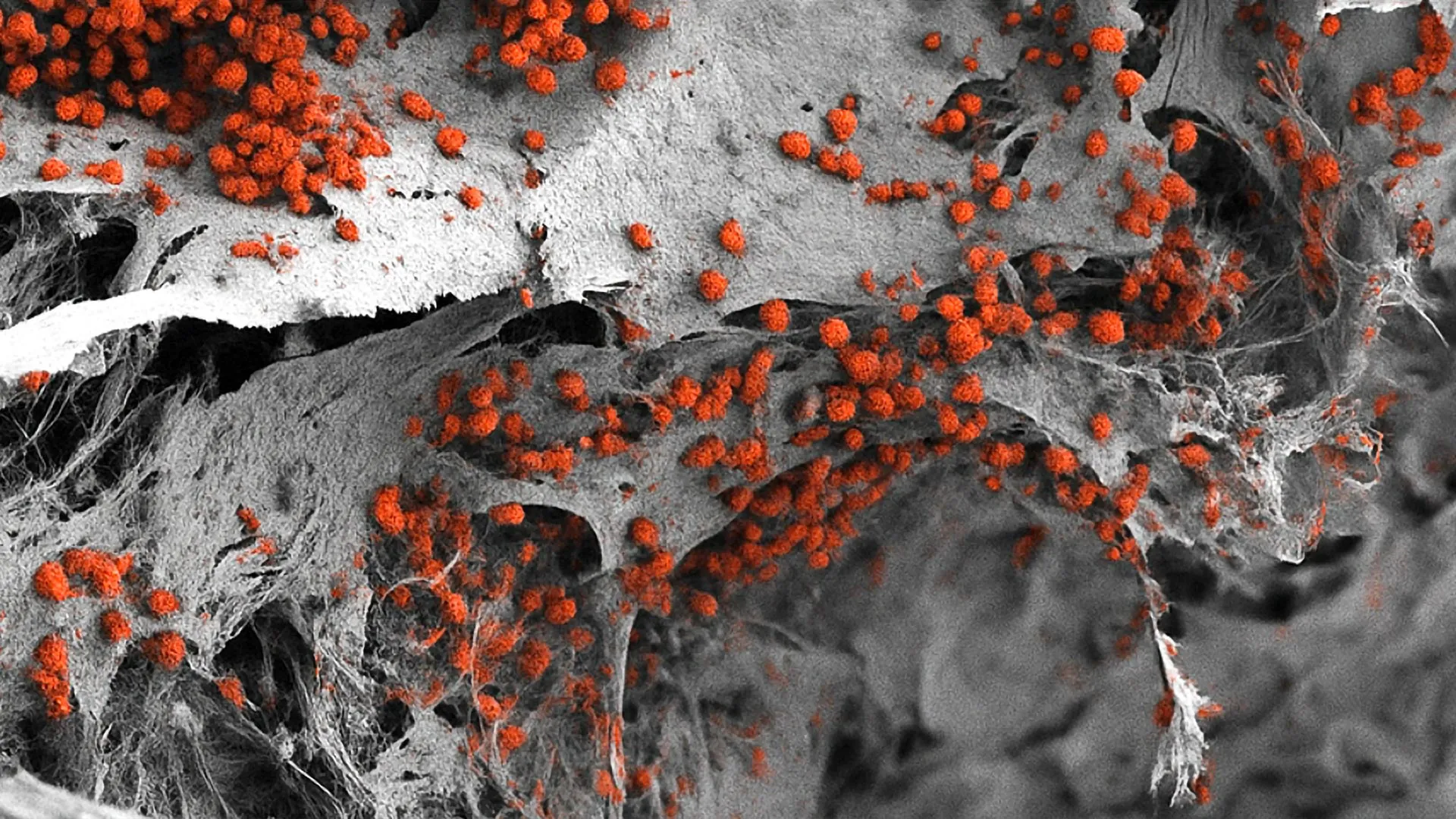
During the first trimester, specifically from the fifth to the eleventh week post-conception, the human embryo undergoes a rapid transformation. The researchers discovered that for most of the skeleton, cartilage acts as a temporary scaffold. This cartilage is gradually replaced by bone cells in a process that requires precise timing and communication between different cell lineages. By capturing this process at a single-cell level, the team was able to describe the gene networks and interaction "checkpoints" that ensure a limb or a vertebra forms correctly.
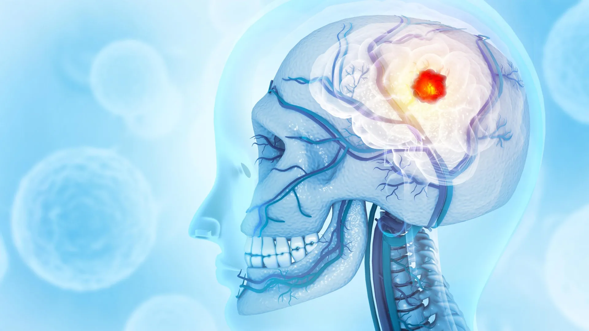
One of the most significant findings involves the "calvarium," or the top of the skull. Unlike the long bones of the arms or legs, the top of the skull does not use a cartilage scaffold. Instead, the researchers identified unique types of early bone cells that form the skull directly. This distinction is vital for understanding why certain genetic mutations affect the skull differently than the rest of the body.
Insights into Craniosynostosis and Skull Development
A primary focus of the study was the investigation of craniosynostosis, a condition affecting approximately one in every 2,500 births. In a typical infant, the skull is composed of several plates joined by "soft spots" or sutures. these sutures allow the skull to remain flexible so the brain can continue its rapid expansion during the first two years of life. Usually, these sutures fully fuse and harden by the age of two.
In cases of craniosynostosis, these sutures fuse prematurely. This prevents the brain from growing outward, leading to an abnormal head shape and, if left untreated, increased intracranial pressure. This pressure can result in permanent brain damage, hearing loss, vision impairment, and significant learning difficulties. While surgeons in the UK and worldwide have developed effective procedures to reopen these sutures, the underlying cellular cause has remained elusive.
By using the new skeletal atlas, the Sanger Institute team identified the specific early bone cells disrupted by the genetic mutations associated with craniosynostosis. For the first time, researchers can see exactly which pathways are "misfiring" to cause early fusion. This discovery opens the door for future diagnostic tools that could identify the risk of craniosynostosis earlier and potentially lead to non-surgical therapeutic interventions that regulate bone growth in the womb or shortly after birth.
The Developmental Roots of Osteoarthritis
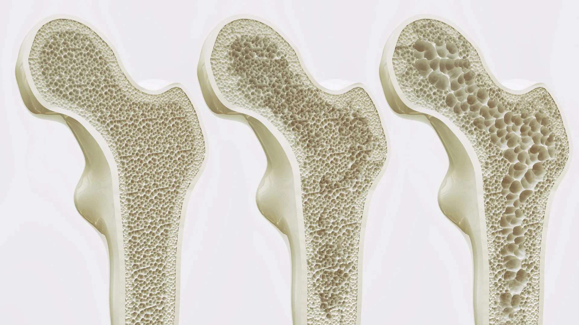
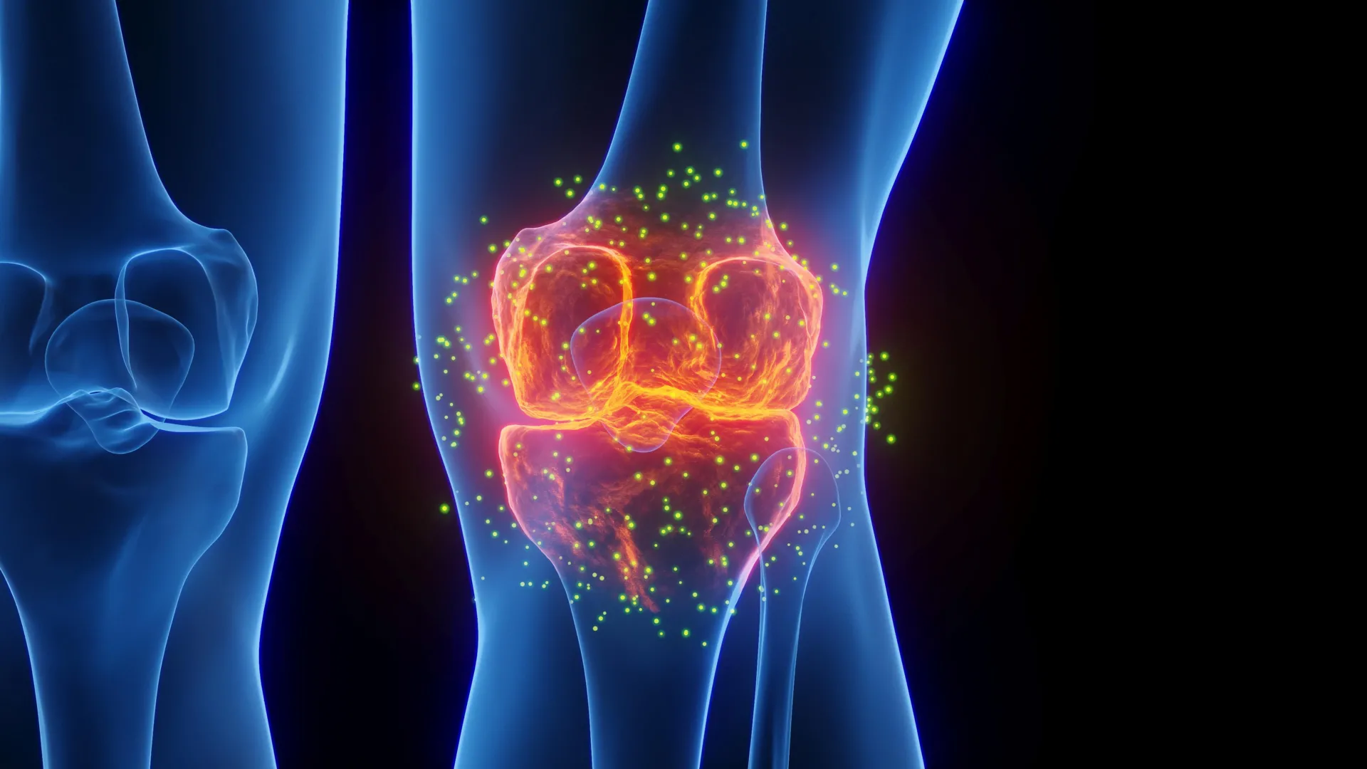
Perhaps the most surprising revelation of the skeletal atlas is its connection to the ageing skeleton. Osteoarthritis (OA) is the most prevalent joint disorder globally, affecting millions of people. In the United Kingdom alone, it is the leading cause of joint pain and stiffness, often requiring total hip or knee replacements when the protective cartilage wears away. Because adults lack the ability to naturally regenerate damaged cartilage, OA is currently managed rather than cured.
The researchers compared the genetic variants known to increase the risk of adult osteoarthritis with the gene expression patterns found in the developing fetal skeleton. They discovered a clear divergence in the origins of hip and knee arthritis. Genetic variants associated with a high risk of hip osteoarthritis were found to be active during the early development of bone cells and their downstream regulators. In contrast, variants linked to knee arthritis were more closely associated with the cells involved in cartilage formation and repair.
This suggests that the "seeds" of arthritis may be sown during the very earliest stages of skeletal formation. If a person’s genetic makeup predisposes them to have slightly different cartilage repair mechanisms or bone growth patterns during gestation, it may dictate how their joints wear down decades later. Understanding these early pathways provides a roadmap for regenerative medicine, as scientists can now look at the "blueprint" of how the body originally built cartilage to find ways to trigger similar repair mechanisms in adults.
Drug Safety and Pharmacological Implications
Beyond disease origins, the skeletal atlas serves as a vital resource for pharmacological safety. Many medications are contraindicated during pregnancy because of their potential to interfere with fetal development. However, the specific mechanisms by which these drugs disrupt growth are often poorly understood.
The research team compiled a list of 65 clinically approved drugs that are currently not recommended for use during pregnancy. By overlaying the known targets of these drugs onto the skeletal atlas, they were able to highlight exactly where and how these therapeutics might disrupt bone and cartilage growth. This creates a "safety map" that pharmaceutical companies and regulatory bodies can use to evaluate the risks of new and existing medications.
This proactive approach to drug safety could be life-changing for pregnant patients who require treatment for chronic conditions. By knowing which cellular pathways are most vulnerable at specific weeks of gestation, doctors can make more informed decisions about which medications are truly dangerous and which might be used in emergencies with minimal risk to the fetal skeleton.
Expert Reactions and Global Collaboration
The release of the atlas has been met with acclaim from the scientific community. Dr. Ken To, a co-first author from the Wellcome Sanger Institute, emphasized the therapeutic potential of the findings. "Having this ‘blueprint’ of bone formation can help us develop effective ways to grow bone and cartilage cells in a dish," Dr. To stated. He noted that characterizing the mechanisms of bone formation allows scientists to predict how genetic changes will impact the skeleton throughout a person’s life.
Dr. Jan Patrick Pett, also a co-first author, highlighted the computational achievement of the project. "Our multi-layered, time- and space-resolved atlas enabled novel computational analyses, which we used to create an integrated view of how developmental processes are regulated," he said. This integrated view is essential for moving from "big data" to "actionable medicine."
Professor Sarah Teichmann, a co-founder of the Human Cell Atlas and senior author of the study, placed the work in the context of the global HCA initiative. "This detailed atlas of bone development in space and time is coordinated with other studies which brings the entire Human Cell Atlas initiative one step closer to fully understanding what happens in the human body across development, health, and disease," Teichmann remarked. She emphasized that the atlas is freely available to the research community worldwide, ensuring that the data can be used to accelerate discoveries in any laboratory.
Chronology of Skeletal Development and Research
To understand the scope of the atlas, it is helpful to look at the timeline of human skeletal growth and how this study intersects with it:
- Weeks 5-11 Post-Conception: The primary window of the Sanger Institute study. This is the period when the basic layout of the skeleton is established via cartilage scaffolds and initial bone cell differentiation.
- First Trimester: The period when the 65 identified "at-risk" drugs can cause the most significant developmental disruptions.
- Birth to Age 2: The period when the soft spots (fontanelles) in the skull typically remain open to allow brain growth. This is the critical window for diagnosing and treating craniosynostosis.
- Adulthood: The phase where the genetic predispositions identified in the atlas manifest as osteoarthritis. The lack of regenerative capacity in adult cartilage makes the "instructions" found in the fetal atlas invaluable for future therapies.
Broader Impact and the Future of Regenerative Medicine
The implications of this skeletal blueprint extend far beyond the specific conditions of arthritis and craniosynostosis. By understanding the "instruction manual" used by the body to build limbs and joints, scientists are moving closer to the reality of lab-grown tissues. For patients with severe bone fractures that won’t heal, or those with degenerative joint diseases, the ability to replicate the embryonic growth environment could lead to biological implants that are far superior to current metal and plastic prosthetics.
Furthermore, the study underscores the importance of the Human Cell Atlas as a foundational resource for modern medicine. Just as the Human Genome Project provided the list of "parts" for human biology, the HCA is providing the "assembly instructions." The skeletal atlas is a primary chapter in that manual, offering hope for a future where bone and joint conditions can be diagnosed before birth and treated with precision throughout a lifetime.
The data is now hosted on a public portal, allowing researchers to explore the gene expression of any cell type in the developing skeleton. As this resource is integrated with other HCA data—such as maps of the developing nervous and circulatory systems—it will provide a holistic view of how the human body builds itself, one cell at a time.