New research emerging from the Fralin Biomedical Research Institute at VTC is fundamentally questioning the established framework used to study and treat chronic neurological conditions, including dystonia, ataxia, and essential tremor. Led by Meike van der Heijden, an assistant professor at the institute and within Virginia Tech’s School of Neuroscience, the study suggests that a long-held assumption regarding the relationship between different types of brain cells may be flawed. For decades, the scientific community has operated under the premise that monitoring the activity of Purkinje cells—the large, accessible neurons on the surface of the cerebellum—provides an accurate proxy for the activity of the deep cerebellar nuclei cells located further within the brain. However, this new evidence, published in the Journal of Physiology, indicates that the correlation between these two cell populations is neither linear nor reliably predictive, potentially necessitating a paradigm shift in how cerebellar diseases are researched and managed.
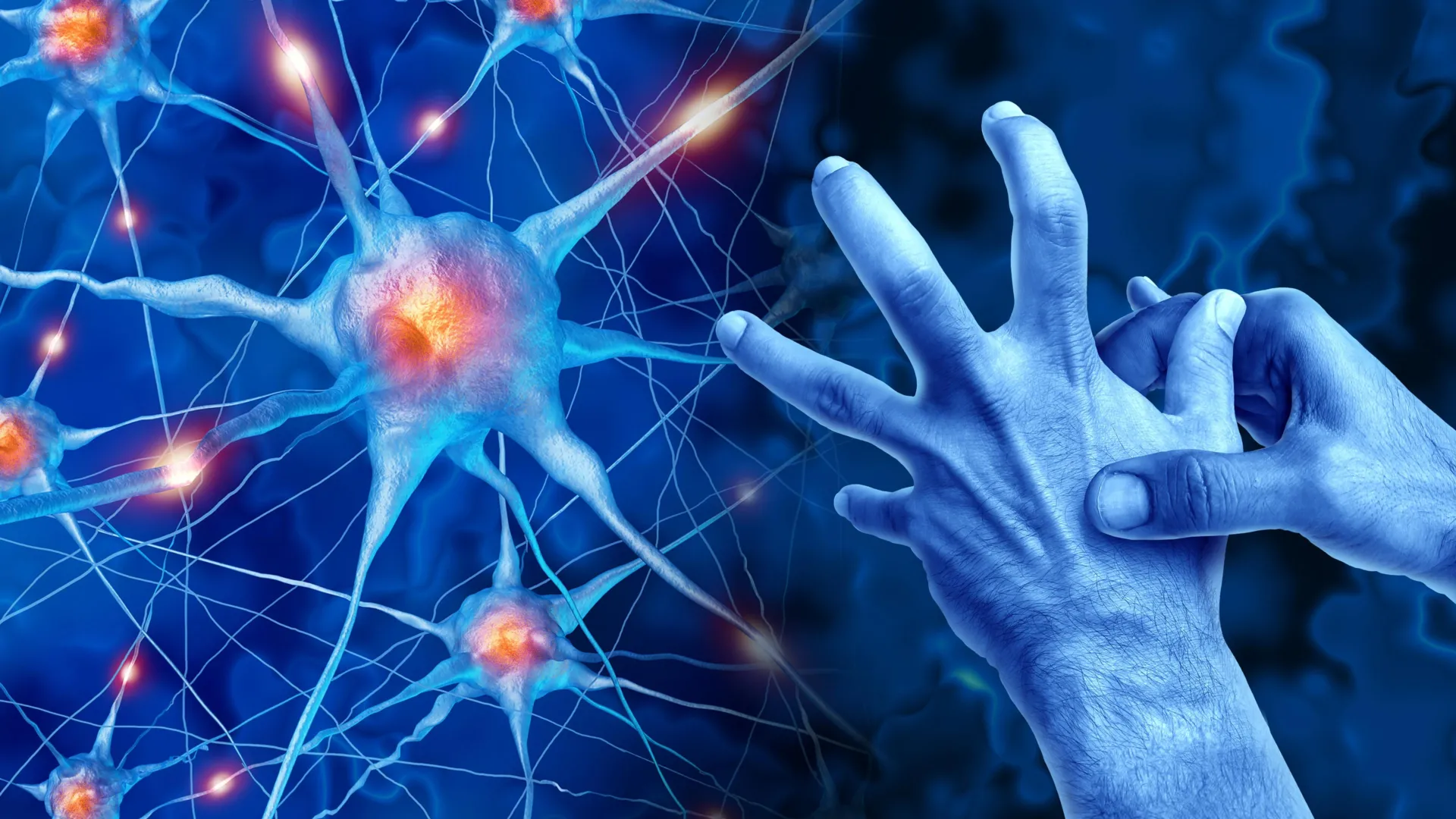
The cerebellum, often referred to as the "little brain," is a complex structure situated at the back of the skull. While it represents only about 10% of the brain’s total volume, it contains more than half of its neurons. Its primary function is the coordination and refinement of motor movements, ensuring they are smooth, timed correctly, and accurate. When the internal communication of the cerebellum is disrupted, the results are often debilitating. Patients may suffer from dystonia, characterized by painful, involuntary muscle contractions; ataxia, which leads to a lack of muscle control and balance; or tremors, involving uncontrollable rhythmic shaking. Understanding the internal "wiring" of this region is therefore critical for developing effective therapies.
The Traditional Model of Cerebellar Communication
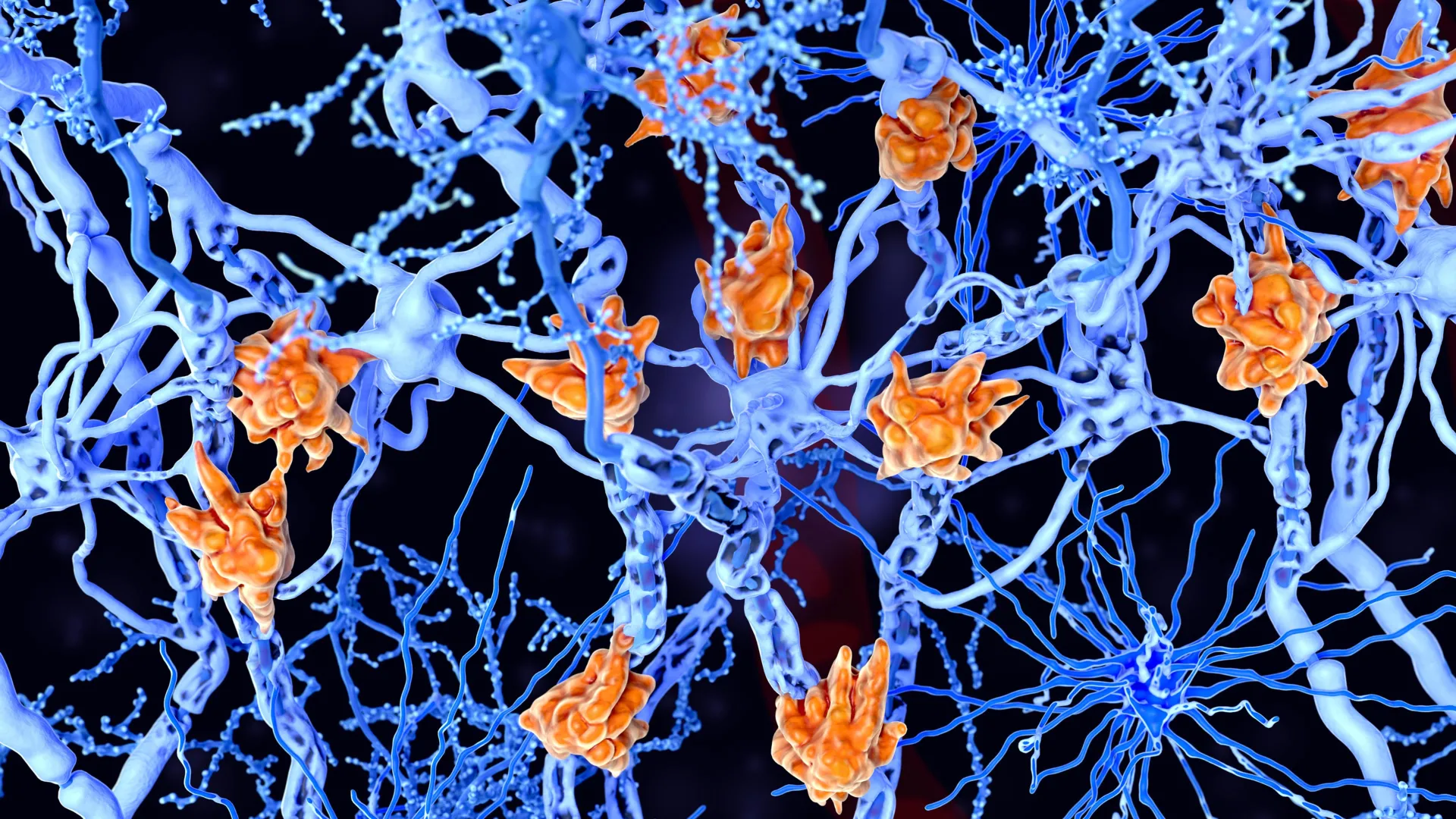
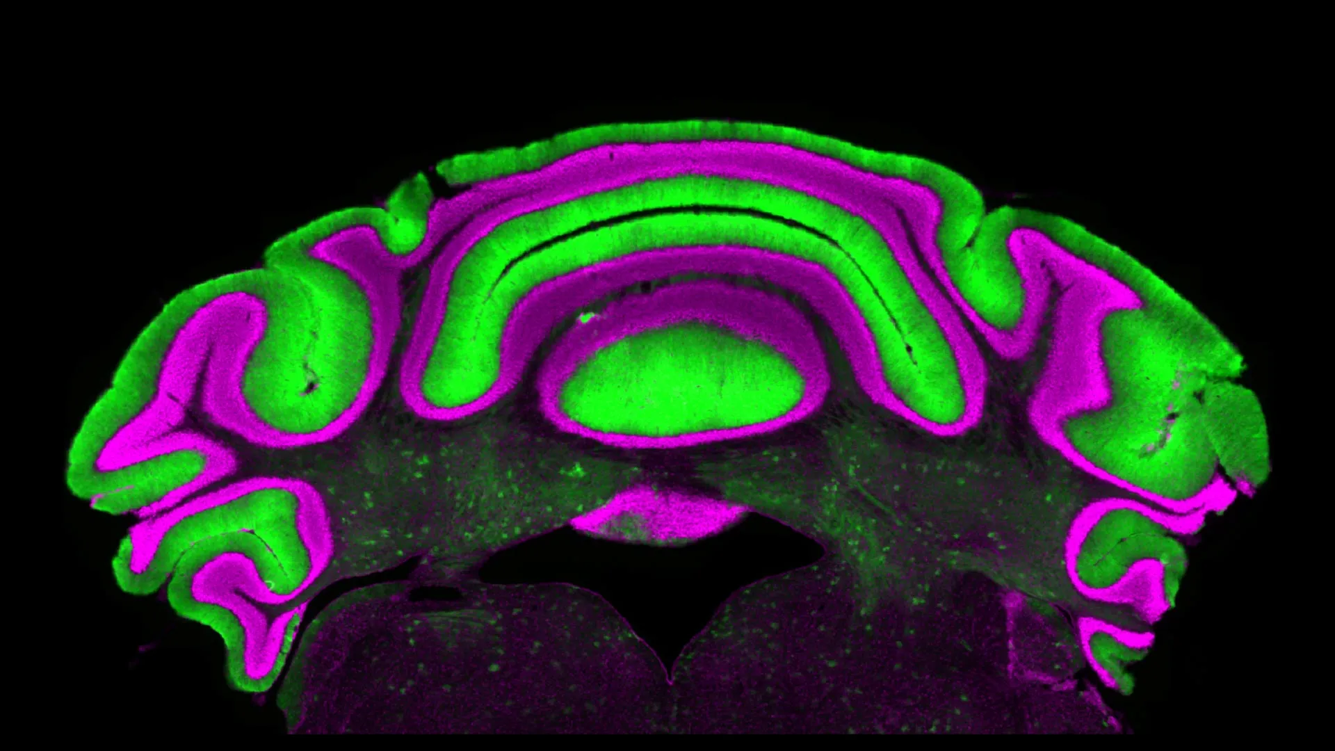
To understand the significance of the Virginia Tech study, one must first look at the traditional neuroscientific model of the cerebellum. The architecture of the cerebellum is organized into layers. On the outer cortex sit the Purkinje cells, which are among the most distinctive neurons in the human body due to their massive, tree-like dendritic structures. These cells serve as the primary output of the cerebellar cortex. Their role is inhibitory; they release a neurotransmitter called GABA (gamma-aminobutyric acid), which suppresses the activity of the cells they target.
Those targets are the deep cerebellar nuclei (DCN) cells. Located deep within the white matter of the cerebellum, the DCN cells are the final output station for the entire cerebellar structure. They send signals out to the rest of the brain and the spinal cord to execute movements. Because Purkinje cells are the direct upstream regulators of the DCN, the scientific consensus has long been that "more activity" in Purkinje cells automatically equates to "less activity" in the deep nuclei, and vice versa.
This assumption was not merely theoretical; it was a practical necessity. Purkinje cells are relatively easy to record because of their size and their location near the surface of the brain. In contrast, the deep nuclei are physically difficult to reach, buried under layers of dense tissue and requiring highly invasive procedures to monitor in real-time. Consequently, researchers have frequently used Purkinje cell firing rates as a "biomarker" or a mirror for what they assumed was happening in the deeper, more influential regions of the cerebellum.
Methodology and Findings: Breaking the Correlation
The research team, led by Van der Heijden and first author Alyssa Lyon, a doctoral candidate in Virginia Tech’s Translational Biology, Medicine, and Health Graduate Program, sought to test the validity of this mirror-image relationship. They moved beyond simple observation and utilized an extensive database of electrophysiology recordings. These recordings were taken from pre-clinical models specifically designed to mimic cerebellar diseases.
Electrophysiology involves the use of microelectrodes to measure the electrical activity (spikes or action potentials) of individual neurons. By comparing the firing patterns of Purkinje cells against the firing patterns of deep nuclei cells across various disease states, the researchers expected to see a clear, inverse correlation. Instead, the data revealed a surprising lack of synchronicity.
"We see that there’s not a clear linear relationship between activity in the Purkinje cells and in the deep nuclei cells," Van der Heijden stated. "So there’s very limited predictive power in monitoring one to understand what’s going on in the other."
The study found that even when Purkinje cells exhibited high levels of irregular activity—a hallmark of many movement disorders—the deep nuclei cells did not always respond in a predictable fashion. In some instances, the DCN cells maintained their own patterns of activity regardless of the inhibitory signals coming from the Purkinje cells. This suggests that the DCN are not merely passive recipients of Purkinje signals but are complex processors that integrate various inputs, some of which may bypass or override the Purkinje pathway.
A Chronology of Cerebellar Understanding
The history of cerebellar research explains why the "Purkinje-centric" view became so dominant.
- Late 19th Century: Johannes Purkinje first identifies the large neurons that now bear his name. Their unique shape makes them the focal point of early neuroanatomical studies.
- Early 20th Century: Santiago Ramón y Cajal maps the circuitry of the cerebellum, identifying the inhibitory link between the cortex and the nuclei.
- 1960s-1970s: The "Marr-Albus-Ito" model of the cerebellum is developed, proposing that the cerebellum acts like a learning computer, with Purkinje cells acting as the primary site of motor memory and error correction.
- 1990s-2010s: The rise of Deep Brain Stimulation (DBS) and optogenetics allows researchers to manipulate specific cell types. However, due to the ease of access, many studies continue to focus on the cerebellar cortex (Purkinje cells) rather than the deep nuclei.
- Present Day: The Virginia Tech study represents a modern "course correction," highlighting that the output of the system (the DCN) cannot be fully understood by looking only at its primary input (the Purkinje cells).
Implications for Dystonia, Ataxia, and Tremor
The disconnect discovered by the Virginia Tech team has profound implications for the clinical treatment of movement disorders. Currently, many therapeutic strategies—ranging from pharmacological interventions to experimental neuromodulation—aim to "fix" the firing patterns of Purkinje cells. The logic has been that if the Purkinje cells return to a normal firing rate, the deep nuclei will follow suit, and the patient’s tremors or muscle contractions will cease.
However, if the relationship is non-linear, a treatment that successfully stabilizes Purkinje cells might have no effect on the deep nuclei, or it might even exacerbate the problem in the DCN.
"Purkinje and cerebellar deep nuclei cell activity is disrupted in a disease state, and a better understanding of the relationship between these neuron types will ultimately help optimize treatments," said Alyssa Lyon. The research suggests that the "target" for future therapies may need to shift deeper into the brain.
For a patient with ataxia, this could mean that current medications are targeting the wrong cell population. For those undergoing Deep Brain Stimulation, it suggests that the placement of electrodes and the frequency of the electrical pulses may need to be recalibrated to account for the independent behavior of the deep nuclei.
Analyzing the Data: Why the Disconnect Exists
While the study proves the lack of correlation, it also opens new questions about why the deep nuclei are so independent. Scientists point to several factors that might explain this:
- Convergence Ratios: In the human brain, hundreds of Purkinje cells may converge onto a single deep nuclei cell. This massive "many-to-one" ratio means that the activity of a single Purkinje cell, or even a small group of them, is easily drowned out by the collective input of the entire population.
- Extracerebellar Inputs: The deep nuclei receive direct excitatory inputs from other parts of the brain, such as the mossy fibers and climbing fibers, which do not pass through the Purkinje cells first. These inputs can stimulate the DCN even when Purkinje cells are trying to inhibit them.
- Intrinsic Pacemaking: Deep nuclei cells have their own internal "pacemakers." They are capable of firing spontaneously even in the absence of external stimuli. This intrinsic rhythmicity may be more resilient to Purkinje interference than previously thought.
A Cautionary Tale for Future Research
The findings serve as a significant warning to the broader neuroscience community. Van der Heijden describes the study as a "cautionary tale," emphasizing the dangers of scientific assumptions. In the world of drug development, many compounds are screened based on their ability to affect the "biomarker"—in this case, Purkinje cell activity. If that biomarker does not actually represent the disease’s functional output, years of research and millions of dollars could be spent on drugs that fail in clinical trials because they are effectively treating the wrong symptom.
"We need to be very careful in making assumptions, and to actually do experiments to test our hypotheses," Van der Heijden noted.
The study advocates for a more holistic approach to cerebellar research. It suggests that future studies must utilize more sophisticated recording techniques that can reach the deep nuclei, despite the technical challenges. Furthermore, it highlights the need for computational models of the brain that can account for the non-linear, complex interactions between cell layers.
Broader Impact and the Path Toward Personalized Medicine
Beyond the specific disorders of dystonia and ataxia, this research touches on the growing field of personalized neurology. As scientists realize that brain circuits do not always follow simple "if-then" rules, the need for patient-specific mapping becomes more apparent. If the Purkinje-DCN relationship varies between individuals or between different stages of a disease, then a "one-size-fits-all" approach to treatment is unlikely to succeed.
The Fralin Biomedical Research Institute’s findings are expected to prompt a re-evaluation of existing datasets in labs across the globe. Researchers may now go back to older studies to see if the "failed" results were actually a result of monitoring the wrong cell type.
In the long term, this research could lead to the development of new diagnostic tools that focus on the deep nuclei. While currently difficult to access, advancements in high-resolution functional MRI (fMRI) and deep-seated electrode arrays may eventually allow clinicians to monitor the DCN as easily as they currently monitor the surface of the brain.
By dismantling a long-held myth in neuroscience, Van der Heijden and her team have not only cleared a path for more accurate research but have also provided a new sense of hope for patients. For those living with the daily challenges of movement disorders, a deeper understanding of the brain’s "little brain" is the first step toward more effective, targeted, and life-changing therapies. The message from Virginia Tech is clear: to truly understand the message the brain is sending, one must listen to the final messenger, not just the intermediary.