Researchers at the Institute for Neurosciences in Spain and the École Polytechnique Fédérale de Lausanne in Switzerland have announced the discovery of an experimental molecule, designated as OLE, which demonstrates the capacity to reprogram the brain’s innate immune system to fight the progression of Alzheimer’s disease. The study, published in the peer-reviewed journal Cell Death and Disease, details how this compound restores the protective capabilities of microglia—cells that act as the primary immune defense within the central nervous system. By shifting these cells from a dysfunctional state back into an active, protective role, the researchers have opened a significant new front in the global effort to treat neurodegenerative disorders.
The collaborative effort was spearheaded by Dr. José Vicente Sánchez Mut, leader of the Functional Epi-Genomics of Aging and Alzheimer’s Disease laboratory at the Institute for Neurosciences (IN), a joint venture of the Spanish National Research Council (CSIC) and Miguel Hernández University of Elche (UMH). He worked in close coordination with Johannes Gräff of the École Polytechnique Fédérale de Lausanne (EPFL). Their findings suggest that the cognitive decline associated with Alzheimer’s is not merely a result of protein accumulation, but also a failure of the brain’s internal "janitorial" system, which OLE appears to successfully reboot.
A Paradigm Shift in Neurodegenerative Research
For decades, Alzheimer’s research has been dominated by the "amyloid hypothesis," which posits that the accumulation of beta-amyloid plaques is the primary driver of the disease. While this remains a central tenet of the field, recent clinical trials of drugs designed solely to clear these plaques have met with mixed success. This has led the scientific community to look more closely at the role of neuroinflammation and the immune environment of the brain.
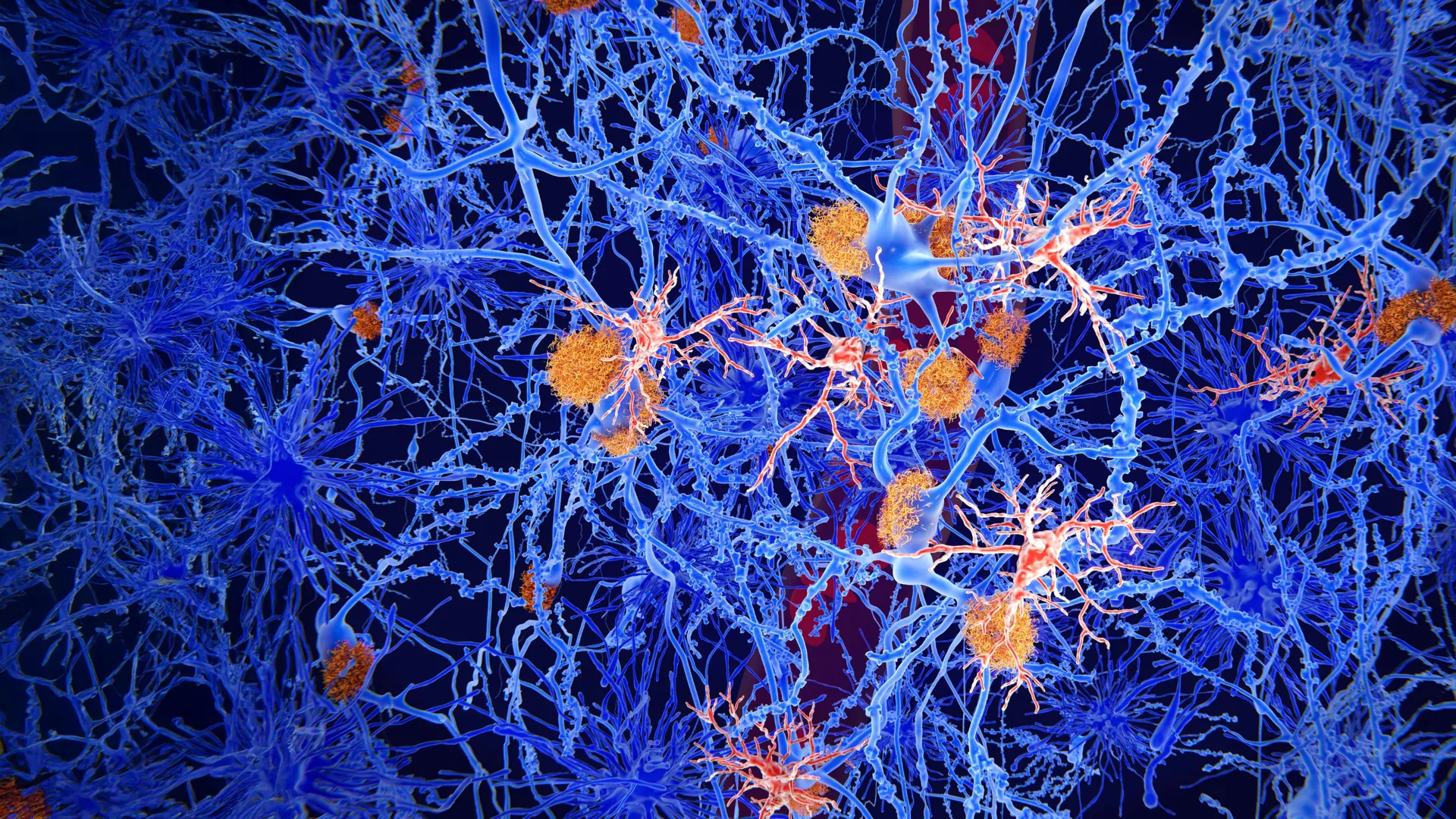
Microglia, which make up about 10% to 15% of all cells found within the brain, are responsible for identifying and neutralizing pathogens, as well as clearing out metabolic waste and dead neurons. In a healthy brain, they are highly mobile and efficient. However, in the presence of Alzheimer’s, these cells undergo a paradoxical transformation. Instead of clearing the beta-amyloid plaques, they become "exhausted" or enter a state of chronic inflammation that further damages the surrounding neurons.
The OLE molecule, derived from the PM20D1 gene, addresses this specific failure. Rather than acting as a simple scavenger of plaques, OLE acts as a biochemical signal that "re-educates" the microglia. This reprogramming enables the cells to regain their motility and their ability to physically sequester toxic aggregates, preventing them from coming into contact with healthy brain tissue.
The Role of the PM20D1 Gene and the Discovery of OLE
The genesis of this research lies in the study of the PM20D1 gene, which has previously been identified as a protective factor against Alzheimer’s in genetic association studies. Researchers observed that individuals with higher expressions of this gene often showed a slower progression of the disease. OLE is a small molecule metabolite associated with this genetic pathway.
Dr. Sánchez Mut and his team hypothesized that if the protective effects of PM20D1 could be distilled into a therapeutic compound, it might provide a way to bolster the brain’s natural defenses even after the onset of the disease. Through a series of complex biochemical screens, OLE emerged as a primary candidate for this intervention. The molecule was found to modulate the metabolic pathways of microglia, essentially giving them the "energy" and the "instructions" needed to resume their protective duties.
Experimental Methodology: From Worms to Mammals
To validate the efficacy of OLE, the research team employed a multi-stage experimental approach involving different biological models. This progression allowed the scientists to observe the molecule’s effects on both simple organisms and complex mammalian systems.
Rapid Assessment in C. elegans
The first phase of the study utilized Caenorhabditis elegans, a species of translucent roundworm frequently used in aging research. These specific worms were genetically modified to produce human beta-amyloid, which leads to rapid protein aggregation and physical paralysis. Because C. elegans has a short lifespan and a well-mapped nervous system, it served as an ideal "first responder" model for testing OLE’s toxicity and basic protective effects.
The results were striking. Worms treated with OLE showed a significant reduction in the accumulation of protein aggregates. More importantly, their motor function—which typically declines as amyloid toxicity takes hold—was preserved. This provided the "proof of concept" necessary to move the research into more complex animal models.
Cognitive Recovery in Murine Models
The second phase involved mouse models specifically bred to exhibit the hallmarks of Alzheimer’s disease, including memory loss and extensive plaque formation. These mice were administered OLE over a three-month period. To measure the impact on cognitive function, the researchers employed a series of memory-based behavioral tests, such as the Morris Water Maze and object recognition tasks.
The data indicated that the treated mice performed significantly better on these tests than the control group. From a physiological standpoint, the brains of the OLE-treated mice showed a different landscape than those of the untreated mice. While plaques were still present, they were smaller and, crucially, were surrounded by a dense "halo" of active microglia.
Precision Mapping: Single-Cell Analysis of the Brain’s Immune Response
To understand exactly how OLE was achieving these results, the team utilized cutting-edge single-cell RNA sequencing. This technology allowed the researchers to examine the gene expression profiles of thousands of individual cells within the mouse brains.
The analysis confirmed that microglia were the primary responders to the OLE treatment. "Single-cell analysis allowed us to determine that microglia were the cells that responded most strongly to the treatment," noted Victoria Pozzi, the study’s first author. The data showed that OLE activated specific genetic pathways associated with lipid metabolism and cell motility.
In essence, OLE acted as a GPS for the immune cells, directing them toward the beta-amyloid deposits. Once the microglia reached the plaques, OLE stimulated the formation of a physical barrier. This "containment strategy" is a novel approach; rather than attempting to dissolve the plaques entirely—which can sometimes release toxic "seeds" of amyloid into the surrounding brain—the OLE-reprogrammed microglia effectively quarantined them.
Containment vs. Clearance: A New Strategy Against Amyloid Toxicity
The discovery of OLE highlights a growing consensus in neurology: that the toxicity of beta-amyloid plaques depends largely on their interaction with neurons. When plaques are "loose" in the brain, they can damage synapses and trigger a cascade of cell death. However, when microglia surround these plaques, they create a protective "insulation" that prevents the toxic surface of the plaque from touching the neurons.
The study’s cell culture experiments further supported this. When neurons were exposed to Alzheimer’s-like conditions in a laboratory dish, the addition of OLE-treated microglia significantly increased neuronal survival rates. This suggests that the molecule’s primary value may lie in its ability to mitigate the collateral damage of the disease, potentially slowing the transition from early-stage impairment to severe dementia.
Intellectual Property and the Path to Clinical Application
The significance of these findings is underscored by the filing of two European patents, one of which is owned by the Spanish National Research Council (CSIC). Patents are a critical step in the "translational" pipeline, as they provide the legal framework necessary for pharmaceutical companies to invest in the multi-million dollar clinical trials required to bring a drug to market.
The researchers emphasize that while the results in animal models are promising, the journey to a human treatment is still in its early stages. The next steps will involve optimizing the delivery of OLE—ensuring it can cross the blood-brain barrier effectively in humans—and conducting safety trials to ensure there are no adverse inflammatory reactions.
"Our results suggest that the impairment of microglia seen in Alzheimer’s is not necessarily permanent," Dr. Sánchez Mut explained. "By identifying a molecule that can reverse this process, we have opened new therapeutic and research avenues that were previously thought to be closed."
Global Context and the Future of Alzheimer’s Therapy
The urgency of this research cannot be overstated. According to the World Health Organization (WHO), more than 55 million people worldwide are currently living with dementia, a figure expected to rise to 139 million by 2050 as the global population ages. Alzheimer’s disease accounts for approximately 60% to 70% of these cases.
Current treatments, such as the recently approved monoclonal antibodies Lecanemab and Donanemab, focus on clearing amyloid from the brain. However, these treatments are expensive, require regular infusions, and carry risks of brain swelling and microhemorrhages. A small-molecule approach like OLE, which focuses on enhancing the brain’s natural immune resilience, could eventually offer a more affordable and potentially safer alternative or a powerful companion therapy.
The study was a massive undertaking supported by a diverse array of international funding bodies, including the Dementia Research Switzerland – Synapsis Foundation, the Pasqual Maragall Foundation, and various Spanish and European government agencies. This level of cross-border cooperation reflects the global nature of the Alzheimer’s crisis and the collective scientific resolve required to address it.
As the research moves toward the next phase of development, the scientific community remains cautiously optimistic. If OLE can replicate its success in human subjects, it may represent a turning point in how we understand and treat the most common form of dementia, shifting the focus from simply fighting the disease to empowering the brain to heal itself.