In a landmark achievement for the field of hematology and regenerative medicine, scientists at the Indiana University School of Medicine have announced the development of a sophisticated imaging methodology designed to visualize the complex interior of bone marrow. This new technique, which utilizes advanced multiplex imaging technology, overcomes long-standing physical and biological hurdles that have historically hindered the study of this critical tissue in mouse models. By providing an unprecedentedly clear view of the bone marrow’s cellular architecture, the research opens new pathways for the development of targeted therapies for a wide spectrum of conditions, ranging from aggressive blood cancers like leukemia to debilitating autoimmune and musculoskeletal disorders.
The breakthrough, recently detailed in the peer-reviewed journal Leukemia, centers on the application of the Phenocycler 2.0 platform. This tool has allowed researchers to map a record-breaking number of cellular markers within intact bone marrow tissue, preserving the spatial relationships between cells that are often lost in traditional analysis methods. The study represents a collaborative effort between various departments at the IU School of Medicine, highlighting the interdisciplinary nature of modern biomedical innovation.
The Complex Architecture of the Bone Marrow Niche
To understand the significance of this technological leap, one must consider the unique biological environment of the bone marrow. Often referred to as the "niche," the bone marrow is the primary site of hematopoiesis—the process by which the body generates new blood cells. It houses hematopoietic stem cells (HSCs), which have the remarkable ability to differentiate into red blood cells, white blood cells, and platelets.
However, studying this environment has proven notoriously difficult for researchers. Bone marrow possesses a gelatinous, semi-fluid consistency but is entirely encased within a rigid, calcified bone matrix. Historically, this has forced scientists into a binary choice: they could either preserve the bone and sacrifice the ability to see the cells clearly, or they could extract the marrow, thereby destroying the delicate spatial organization of the tissue.
"Bone marrow is difficult to study because it is gelatinous and encased in hard bone," explained Sonali Karnik, PhD, an assistant research professor of orthopedic surgery at the IU School of Medicine and a co-lead author of the study. Dr. Karnik noted that because the marrow is the staging ground for the immune system and the reservoir for valuable stem cells, a non-disruptive imaging approach is essential for understanding how these cells interact with their environment during both health and disease.
Limitations of Traditional Diagnostic and Research Tools
Before the implementation of this new multiplex imaging technique, researchers primarily relied on two methods: flow cytometry and standard immunofluorescence imaging. While both have been instrumental in the progress of modern medicine, they possess inherent limitations that the IU team sought to address.
Flow cytometry is the current gold standard for quantifying cell populations. It involves breaking down a tissue sample into a single-cell suspension and passing those cells through a laser. While this provides highly accurate data on the types of cells present, it completely eliminates the "spatial context." In a disease like leukemia, knowing how many cancer cells are present is important, but knowing exactly where they are located in relation to blood vessels, nerves, or healthy stem cells is arguably more critical for understanding disease progression and treatment resistance.
Standard fluorescence imaging, on the other hand, allows for the visualization of cells within the tissue. However, it is typically limited by the "color barrier." Most standard microscopes can only distinguish between three or four different fluorescent markers at one time. Given that the bone marrow contains dozens of different cell types—including various stages of myeloid and lymphoid cells, endothelial cells, and osteoblasts—a four-marker limit provides only a narrow, "keyhole" view of the total environment.
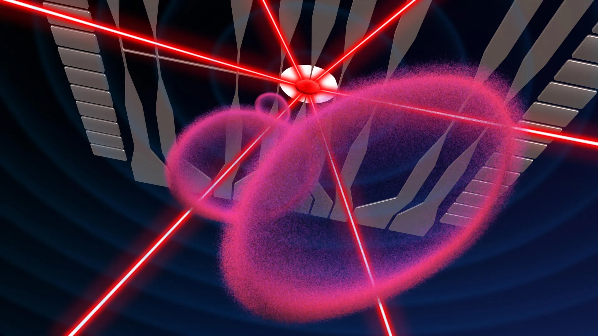
The Phenocycler 2.0: A Paradigm Shift in Spatial Biology
The IU research team overcame these limitations by successfully adapting the Phenocycler 2.0 system for use in bone marrow. The Phenocycler (formerly known as CODEX) utilizes a process of iterative staining and imaging. By using antibodies conjugated to unique DNA barcodes, the system can apply dozens of markers to a single tissue slice.
In this specific study, the researchers were able to visualize 25 different cellular markers simultaneously within intact mouse bone marrow. This allowed them to create a high-resolution, multi-layered map of the tissue. By seeing 25 markers at once, scientists can identify not just the cell types, but also their functional states, their proximity to one another, and how they cluster around specific anatomical features like the inner lining of the bone (the endosteum) or the central blood vessels.
While the Phenocycler technology had previously been utilized to map "softer" organs such as the spleen, liver, and kidneys, the IU Cooperative Center of Excellence in Hematology (CCEH) is the first group to successfully apply the technique to the challenging environment of the mouse bone marrow. This required specialized protocols for tissue preparation that ensure the marrow remains fixed and stable despite the removal or thinning of the surrounding bone.
Chronology of Development and Institutional Collaboration
The development of this technique is the result of several years of focused research within the IU School of Medicine’s ecosystem. The project was spearheaded by the Herman B Wells Center for Pediatric Research, a facility known for its work in developmental biology and childhood blood cancers.
The timeline of the research reflects a steady progression from proof-of-concept to high-dimensional application:
- Initial Phase: Researchers identified the need for better spatial mapping to understand why certain leukemia treatments failed despite high "cell kill" rates in lab settings.
- Technology Acquisition: The IU School of Medicine integrated the Phenocycler 2.0 platform into its core facilities, providing the necessary hardware for multiplexing.
- Protocol Refinement: Over several months, the team, led by Dr. Karnik and co-senior author Reuben Kapur, PhD, refined the chemical processes required to stain bone marrow without degrading the tissue’s structural integrity.
- Data Validation: The team cross-referenced their 25-marker imaging data with traditional flow cytometry results to ensure that the new method was as accurate as established standards.
- Publication and Patenting: Following the successful validation, the findings were submitted to Leukemia, and the IU Innovation and Commercialization Office moved to protect the intellectual property by filing a provisional patent.
Broad Implications for Oncology and Beyond
The ability to see the "neighborhood" of a cell has profound implications for the treatment of leukemia and other hematologic malignancies. Cancer cells do not exist in a vacuum; they often "hijack" the bone marrow niche to protect themselves from chemotherapy. By using this new imaging technique, researchers can identify exactly where these "refugee" cancer cells hide, allowing for the development of drugs that can flush them out of their protective environments.
Reuben Kapur, PhD, director of the Herman B Wells Center for Pediatric Research and co-senior author, emphasized the translational potential of the work. "Because mouse models are widely used to study human diseases, this technique offers a promising new method for investigating a range of conditions like autoimmune diseases, leukemia and other disorders involving bone marrow," Kapur stated.
The impact extends beyond oncology:
- Autoimmune Diseases: In conditions like lupus or rheumatoid arthritis, the bone marrow can become a site of inflammatory cell production. This imaging allows scientists to track the "misfiring" of immune cell development.
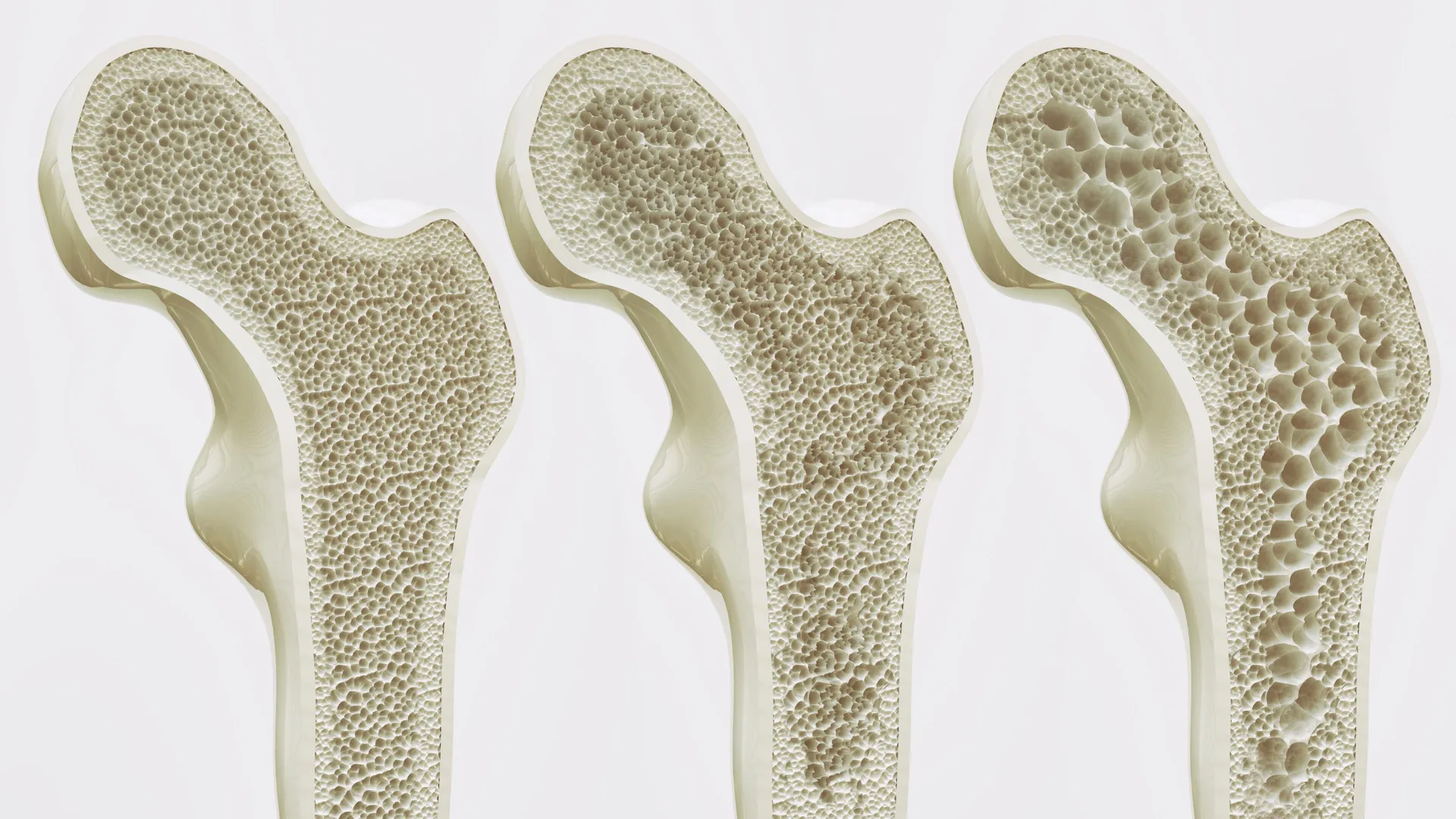
- Musculoskeletal Disorders: As the body ages, the balance between bone-building cells (osteoblasts) and bone-resorbing cells (osteoclasts) shifts. Mapping the marrow-bone interface could lead to new treatments for osteoporosis.
- Stem Cell Transplantation: Improving the success rates of bone marrow transplants requires a deep understanding of how donor cells "home" to the marrow and engraft. This technique provides a literal roadmap for that process.
Supporting Data and Future Technical Expansions
The 25-marker panel used in the published study included markers for various lineages of blood cells, including hematopoietic stem cells, progenitor cells, and mature immune cells. However, the IU team views this as only the beginning. The methodology is designed to be modular, meaning new markers can be added as research needs evolve.
Current efforts are underway to expand the panel to include non-hematopoietic elements. The team is working to integrate markers for:
- Nerve Fibers: To study how the nervous system regulates blood cell release.
- Muscle Cells: To investigate the interplay between physical activity and marrow health.
- Signaling Proteins: To visualize the "chemical messages" being sent between cells in real-time.
Furthermore, the data generated by this technique is massive, often requiring advanced computational tools and artificial intelligence to analyze the millions of data points regarding cell coordinates and marker intensity. This "spatial proteomics" approach is expected to become a cornerstone of the burgeoning field of precision medicine.
Official Support and Collaborative Contributions
The success of this project was made possible through significant institutional and federal support. The research was funded by the National Institutes of Health (NIH), reflecting the federal government’s interest in advancing high-dimensional imaging for human health.
The study also featured a diverse list of contributors from the IU School of Medicine, including Connor Gulbronson, Paige C. Jordan, Rahul Kanumuri, Baskar Ramdas, Ramesh Kumar, Melissa L. Hartman, Izza Khurram, Drew M. Brown, Karen E. Pollok, Pratibha Singh, and Melissa A. Kacena. This extensive list of authors underscores the complexity of the project, which required expertise in surgery, pathology, immunology, and data science.
As the IU Innovation and Commercialization Office moves forward with the patenting process, the methodology is expected to be shared with the broader scientific community through the IU Cooperative Center of Excellence in Hematology. This will likely spark a new wave of research across the globe, as other laboratories adopt these protocols to investigate their own specific disease models.
Conclusion: A New Era of Hematological Discovery
The development of this multiplex imaging technique marks a significant milestone in our ability to decode the mysteries of the bone marrow. By bridging the gap between high-detail cellular analysis and the preservation of anatomical structure, Indiana University researchers have provided the scientific community with a powerful new lens through which to view human disease.
As this technology matures and the marker panels expand, the transition from mouse models to clinical applications in human patients becomes increasingly tangible. For patients suffering from leukemia, bone marrow failure, or chronic inflammation, the clarity provided by this 25-marker "map" may eventually lead to the more precise, more effective, and less toxic therapies of tomorrow.