Antibody-drug conjugates (ADCs) have fundamentally transformed the landscape of modern oncology, evolving from a theoretical "magic bullet" concept into a cornerstone of precision medicine. With a projected market valuation of $57 billion by 2032, according to Evaluate Pharma, the class has seen rapid adoption, now encompassing approximately 15 FDA-approved products. Despite this clinical momentum, the industry faces a persistent paradox: a significant percentage of patients whose tumors test positive for specific surface targets fail to respond to ADC therapy. To address this, Gilead Sciences has entered a strategic collaboration with Nucleai, a leader in AI-powered spatial biology, to decode the complex, often invisible, mechanisms behind treatment resistance.
The ADC Paradox: Beyond Simple Target Expression
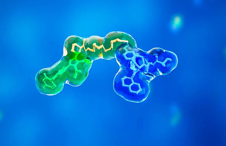
The foundational premise of an ADC is elegant: a monoclonal antibody acts as a homing beacon, delivering a highly potent cytotoxic payload directly to the tumor cell while sparing surrounding healthy tissue. Ideally, this process maximizes therapeutic efficacy while minimizing the systemic toxicities associated with traditional chemotherapy. However, clinical reality is frequently more complex. As Dr. Ken Bloom, head of pathology at Nucleai, observes, the process is fraught with biological hurdles that standard diagnostic assays often fail to capture.
Current pathology standards, primarily Immunohistochemistry (IHC), are designed to identify the presence of a target protein. Yet, an IHC result does not necessarily indicate whether a tumor cell is "vulnerable" to a specific ADC. Research has identified several primary mechanisms of resistance: the therapeutic target may be physically inaccessible due to conformational changes, the extracellular domain of the target may be cleaved by proteases in the tumor microenvironment, or cancer cells may utilize drug-efflux pumps to expel the payload once it enters the cell.
Chronology of a Strategic Partnership
The collaboration between Gilead and Nucleai represents a multi-year progression from exploratory research to deep clinical data integration. The relationship began several years ago, focusing on preclinical multiplex immunofluorescence studies—an approach that allowed for the simultaneous visualization of multiple markers within the tumor microenvironment.
As the partnership matured, the scope expanded to include the retrospective analysis of clinical trial datasets. On August 11, the companies officially disclosed that Nucleai had been analyzing Hematoxylin and Eosin (H&E) and IHC whole-slide images across multiple Gilead oncology indications. By linking granular tissue features observed in these images with actual patient clinical outcomes, the companies are attempting to build a predictive model that transcends traditional biomarker quantification.
The "Inside-Outside" Epitope Problem
One of the most critical technical challenges in ADC development is the discrepancy between what pathologists see and what the drug sees. A notable example is the HER2 receptor. The most common diagnostic assay, the Ventana 4B5 IHC, targets the intracellular domain of the HER2 protein. This allows for reliable detection of the receptor, but it ignores the extracellular domain—the very region that a therapeutic agent like trastuzumab must bind to in order to initiate internalization.
In many tumors, proteolytic shedding or alternative splicing can result in fragments like p95HER2, which lack the extracellular binding site. Consequently, a patient may be categorized as HER2-positive by a pathologist, yet their tumor may be effectively "invisible" to the ADC. This "inside-outside" mismatch is a primary focus of the Gilead-Nucleai analysis, as they seek to move toward diagnostic assays that reflect the actual pharmacological accessibility of the target.
Computational Pathology and Spatial Intelligence
The shift from digital pathology—which primarily digitizes existing workflows—to computational pathology represents a significant leap in diagnostic capability. Computational pathology leverages AI to extract complex spatial relationships that are invisible to the human eye.

"Now we care about the neighborhood that a cell is in," Dr. Bloom noted. "What’s the relationship between it and other cells in the vicinity? What’s the density of things?"
By analyzing the spatial architecture of the tumor microenvironment, the Nucleai platform can identify features that correlate with drug sensitivity. This includes analyzing the density of immune infiltrates, the proximity of tumor cells to vascular structures, and the presence of stromal barriers that may prevent an ADC from reaching its target. These spatial biomarkers provide a multidimensional view that standard, linear IHC scores cannot capture.
Official Perspectives and Translational Impact
For Gilead Sciences, this collaboration is an extension of its commitment to advancing translational medicine. According to Meghna Das Thakur, senior director of oncology biomarkers at Gilead, the partnership combines Nucleai’s expertise in AI-driven spatial biology with Gilead’s deep clinical and scientific infrastructure. The goal is not merely to optimize existing drugs, but to increase the efficiency of biomarker discovery, ensuring that the right patient receives the right therapy at the right time.
Industry analysts suggest that this type of "AI-in-the-loop" model will become the new standard for late-stage clinical trials. By utilizing AI as an "overreader," companies can reduce the inherent variability caused by human fatigue, training differences, and subjective interpretation. This consistency is essential when dealing with subtle markers that might only be predictive of response in specific, localized tissue niches.
Future Implications for Oncology
The implications of this research extend far beyond the current Gilead portfolio. As the industry moves toward more personalized treatment regimens, the ability to predict resistance before a patient receives their first infusion will be paramount. If the Gilead-Nucleai team successfully identifies and validates these new biomarkers, the clinical impact could be profound. It would likely necessitate a shift in how regulatory bodies and clinical pathologists categorize tumor eligibility for ADC therapies.
However, the path to widespread adoption is not without friction. Dr. Bloom points out that while several major pharmaceutical organizations are exploring these methods, a standardized, industry-wide pathway has not yet been established. The current challenge lies in ensuring that these AI-derived measurements are stable, reproducible, and scalable across different laboratory settings globally.
"A fool with a tool is still a fool," Bloom remarked, emphasizing that the future of pathology relies on the marriage of advanced computational power with the seasoned judgment of a trained physician. The goal is not to replace the pathologist, but to provide them with a high-resolution, data-driven map of the tumor landscape.
A Turning Point for Precision Medicine
As the data from this collaboration continues to be analyzed, the industry anticipates that peer-reviewed publications will emerge within the next 6 to 12 months. These findings are expected to shed light on whether current biomarker definitions are sufficient or if a more nuanced, spatially-aware classification system is required for the next generation of ADC therapies.
The partnership underscores a broader transition in drug development: moving away from the "one-size-fits-all" biomarker approach toward a more dynamic understanding of the tumor as an ecosystem. By addressing the physical, structural, and spatial barriers to treatment, Gilead and Nucleai are attempting to solve one of the most stubborn puzzles in modern oncology. If they succeed, they will not only improve patient outcomes for those currently struggling with resistant tumors but also set a new standard for how data-driven pathology informs the future of precision medicine. The race to decode the microenvironment has officially entered a new, computational phase, and the results could define the next decade of cancer treatment.