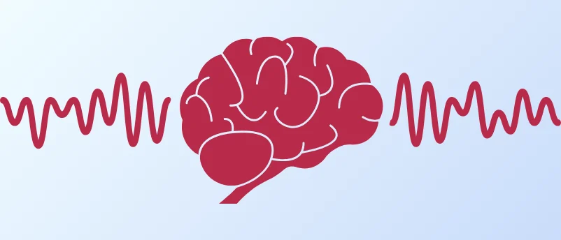
Researchers at the University of Delaware (UD), in collaboration with Nemours Children’s Health, are harnessing the power of artificial intelligence (AI) to revolutionize the early diagnosis of epilepsy, a chronic neurological disorder that affects millions worldwide. Their groundbreaking work focuses on identifying subtle, often imperceptible, patterns within routine brain-wave recordings (electroencephalograms, or EEGs) that traditional diagnostic methods frequently miss, particularly when a seizure is not actively occurring. This innovative approach promises to significantly reduce diagnostic delays, alleviate patient anxiety, and pave the way for more precise and timely therapeutic interventions.
The diagnostic journey for epilepsy is often fraught with uncertainty. While EEGs are a cornerstone of diagnosis, routine recordings typically capture only a brief snapshot of brain activity, usually lasting around 20 to 40 minutes. Given the episodic nature of seizures, it is common for patients not to experience an event during this narrow window, leaving clinicians without the direct observation critical for a definitive diagnosis. In such scenarios, neurologists must rely on interictal abnormalities—subtle electrical disturbances that occur between seizures—which can be exceedingly difficult for the human eye to detect. This challenge often leads to prolonged diagnostic odysseys, causing considerable stress for patients and their families, and potentially delaying access to effective treatment.
The Pressing Need for Improved Epilepsy Diagnostics
Epilepsy is a severe global health concern, affecting approximately 50 million people worldwide, according to the World Health Organization (WHO). In the United States alone, the Centers for Disease Control and Prevention (CDC) estimates that about 3.4 million individuals, including 470,000 children, live with epilepsy. Despite its prevalence, diagnosing epilepsy remains complex. The condition is characterized by recurrent, unprovoked seizures, which are sudden, uncontrolled disturbances in brain activity that can alter consciousness, movement, or sensation. Diagnosis typically involves a detailed medical history, a neurological examination, and an EEG to measure electrical activity in the brain. Magnetic resonance imaging (MRI) and computed tomography (CT) scans may also be used to rule out underlying structural brain abnormalities.
The limitations of standard EEG testing are particularly pronounced in pediatric cases, where children may present with diverse seizure types and less overt symptoms. The inability to capture a seizure during a routine EEG can lead to repeated tests, prolonged monitoring, and a frustrating period of watchful waiting for families. This diagnostic gap not only delays the initiation of appropriate medication or other therapies but also contributes significantly to the psychological burden on patients and caregivers, who live with the constant anticipation of unpredictable seizures. The potential for misdiagnosis or delayed diagnosis underscores the urgent need for more sensitive and objective diagnostic tools.
AI as a "Language Learner" for the Brain
The core innovation developed by the University of Delaware team, led by Austin Brockmeier, an assistant professor in electrical and computer engineering and computer and information sciences, lies in its novel application of machine learning. Brockmeier describes the algorithm’s function as akin to a language learner acquiring an unfamiliar tongue. Instead of relying on predefined patterns or human interpretation of waveforms, the AI is designed to autonomously identify frequently occurring electrical patterns within EEG recordings and learn their contextual significance. This process effectively allows the algorithm to build its own "dictionary" of brainwave patterns, discerning nuances that are too subtle or complex for manual review by human clinicians.
"Our machine-learning approach lets the algorithm learn the brain’s ‘language’ of waveforms, spotting subtle patterns humans might miss during manual review," Brockmeier explained. This capability represents a significant leap beyond traditional EEG analysis, which often relies on visual inspection for hallmark epileptic spikes or sharp waves. The AI’s ability to process vast quantities of data and detect intricate correlations promises a level of diagnostic precision previously unattainable.
The interdisciplinary collaboration that fueled this research began when Brockmeier, a faculty mentor in UD’s interdisciplinary neuroscience graduate (ING) program, presented his computational neuroscience research at an ING seminar. His work immediately captured the attention of Amanda Hernan, an affiliated associate professor of psychological and brain sciences and biomedical engineering at UD and a senior research scientist at Nemours Children’s Health. Hernan, also an ING faculty mentor, specializes in understanding how variations in brain activity impact thinking and learning in children with epilepsy, making her an ideal partner for translating computational insights into clinical applications.
From Mouse Model to Clinical Reality: A Chronology of Discovery
The research unfolded in a structured, phased approach, beginning with a robust proof-of-concept study in animal models before advancing to human trials.

Phase 1: Validation in a Mouse Model (Proof-of-Concept)
The initial phase of the research focused on testing the machine learning algorithm’s efficacy using EEG data from mice engineered with epilepsy-causing variations in the TSC1 gene. The TSC1 gene is implicated in tuberous sclerosis complex (TSC), a genetic disorder that often leads to epilepsy in humans. This model provided a controlled environment to validate the AI’s ability to detect genetic predispositions to epilepsy.
- Study Design: The researchers utilized a diverse panel of more than 40 mice, which included animals with and without the TSC1 gene variation, across three distinct genetic backgrounds or strains. This breadth in genetic backgrounds was crucial for assessing the algorithm’s robustness and generalizability.
- Data Acquisition: For each mouse, EEG segments were meticulously extracted from five days of continuous recordings. Critically, the analysis focused exclusively on segments that contained no visible seizure activity. This deliberate choice forced the algorithm to identify differences solely based on the brain’s baseline electrical activity, mimicking the real-world challenge of diagnosing epilepsy in the absence of an observed seizure.
- Key Findings: The AI algorithm demonstrated remarkable accuracy in distinguishing between the mouse strains and, more importantly, in detecting the TSC1 gene variation in two of the three strains. This outcome was significant because it provided compelling evidence that EEG patterns harbor measurable signals of neurological differences indicative of epilepsy, even when no overt seizures are present.
- Implication: "These results show that EEG patterns contain measurable signals of neurological differences, even without visible seizures," Hernan affirmed. This finding laid the essential groundwork, demonstrating the AI’s potential to uncover previously hidden diagnostic biomarkers.
Phase 2: Transition to Pediatric Clinical Trials
Building on the success of the mouse study, the team is now poised to translate their method into a clinical setting. With vital funding secured from the Delaware Clinical and Translational Research ACCEL Program, Brockmeier and Hernan will apply their AI-driven approach to EEG recordings from children undergoing evaluation for epilepsy at Nemours Children’s Health.
This transition to human pediatric EEGs introduces new complexities. Pediatric recordings are typically shorter than the multi-day recordings used in the mouse study, and children present with a wider spectrum of epilepsy types, each with potentially unique electrical signatures. However, the researchers remain optimistic about the algorithm’s adaptability and potential.
"The goal is to identify biomarkers that flag underlying changes in the brain’s electrical activity before seizures occur," Hernan elaborated. Achieving earlier detection promises to significantly improve patient outcomes by allowing for earlier treatment initiation and reducing the prolonged period of uncertainty and anxiety that families currently endure.
Broader Impact and Future Horizons
The implications of this research extend far beyond mere diagnostic efficiency. For families grappling with the daily unpredictability of epilepsy, earlier and more accurate diagnosis could be life-altering. Hernan poignantly noted the psychological toll of this uncertainty: "Seizures follow natural cycles, but without a way to know where you are in that cycle, the anticipation can be incredibly anxiety-provoking." Reducing this diagnostic limbo would profoundly improve the quality of life for both patients and their caregivers.
Moreover, improved pattern recognition facilitated by AI could dramatically enhance treatment decisions. In current practice, if a new medication is introduced during a natural lull in a patient’s seizure activity, its perceived benefits might be overestimated, leading to suboptimal therapeutic strategies. An AI-powered system could provide a more objective assessment of baseline brain activity, allowing clinicians to make more informed choices about medication efficacy and dosage. This move towards data-driven, personalized treatment aligns perfectly with the principles of precision medicine.
Looking further into the future, the researchers envision a transformative landscape for epilepsy management. One promising avenue involves the development of wearable EEG devices that could provide continuous, real-time monitoring for individuals at high risk of seizures. Such devices, powered by AI algorithms, could offer proactive alerts, enable timely interventions, and provide an unprecedented level of insight into an individual’s seizure patterns and triggers. This continuous data stream could also be invaluable for refining treatment plans over time, leading to more stable and effective seizure control.
The potential applications of this AI methodology are not limited to epilepsy. Brockmeier suggests that similar approaches could eventually be applied to other complex neurological conditions characterized by subtle brainwave abnormalities, such as autism spectrum disorder (ASD) and attention-deficit/hyperactivity disorder (ADHD). The ability to identify precise neurobiological markers for these conditions could unlock new diagnostic pathways and inform targeted therapeutic strategies.
"This is a step toward precision medicine," Brockmeier stated. "Brain-wave typing could help identify which interventions will work best for a given patient." This concept of "brain-wave typing"—characterizing an individual’s unique electrical brain patterns—holds the promise of moving away from a one-size-fits-all approach to neurological care towards highly individualized, patient-specific interventions.
The integration of AI into neurological diagnostics represents a pivotal moment in healthcare. By augmenting human capabilities with advanced computational power, researchers are not only addressing a critical unmet need in epilepsy diagnosis but also laying the groundwork for a future where neurological conditions are understood, diagnosed, and treated with unprecedented precision. For the millions affected by epilepsy, and potentially other neurological disorders, this kind of precision could indeed make a monumental difference, offering hope for earlier intervention, better management, and ultimately, improved lives.