
A team of researchers at the University of Turku in Finland has released a groundbreaking study that fundamentally challenges the prevailing scientific consensus regarding the cause of "brain fog" and other neurological symptoms associated with long COVID. Published in the Journal of Neurology, the study found no evidence of widespread, persistent brain inflammation in individuals suffering from long-term symptoms following a SARS-CoV-2 infection. Instead, the research points toward a complex shift in brain activity, specifically within regions responsible for emotional regulation, memory, and stress responses, suggesting that the physiological underpinnings of the condition may be more nuanced than previously theorized.
Since the early stages of the global pandemic, long COVID—clinically referred to as Post-Acute Sequelae of SARS-CoV-2 (PASC)—has remained one of the most significant and mysterious challenges for modern medicine. Affecting millions of people worldwide, the condition is characterized by a range of debilitating symptoms including chronic fatigue, cognitive impairment (often described as "brain fog"), anxiety, depression, and sleep disturbances. For years, the leading hypothesis among the scientific community was that these symptoms were the result of chronic, low-grade neuroinflammation triggered by the initial viral infection. This new evidence from Finland, however, suggests that while inflammation may play a role in the acute phase, it does not appear to be the primary driver of symptoms in the chronic stage for most patients.
Methodology and Comparative Framework
The study, led by Professor of Neuroimmunology Laura Airas, utilized a sophisticated multi-modal imaging approach to peer into the living brains of those affected. The research team recruited three distinct groups for comparison: 14 individuals diagnosed with long COVID who continued to suffer from neurological symptoms, 11 healthy volunteers who served as the control group, and 13 patients with multiple sclerosis (MS).
The inclusion of MS patients was a critical methodological choice. Multiple sclerosis is a well-characterized autoimmune disease known for causing significant and visible neuroinflammation. By comparing long COVID patients against both healthy individuals and those with a known inflammatory neurological condition, the researchers were able to establish a clear benchmark for what "widespread brain inflammation" actually looks like on a cellular level.
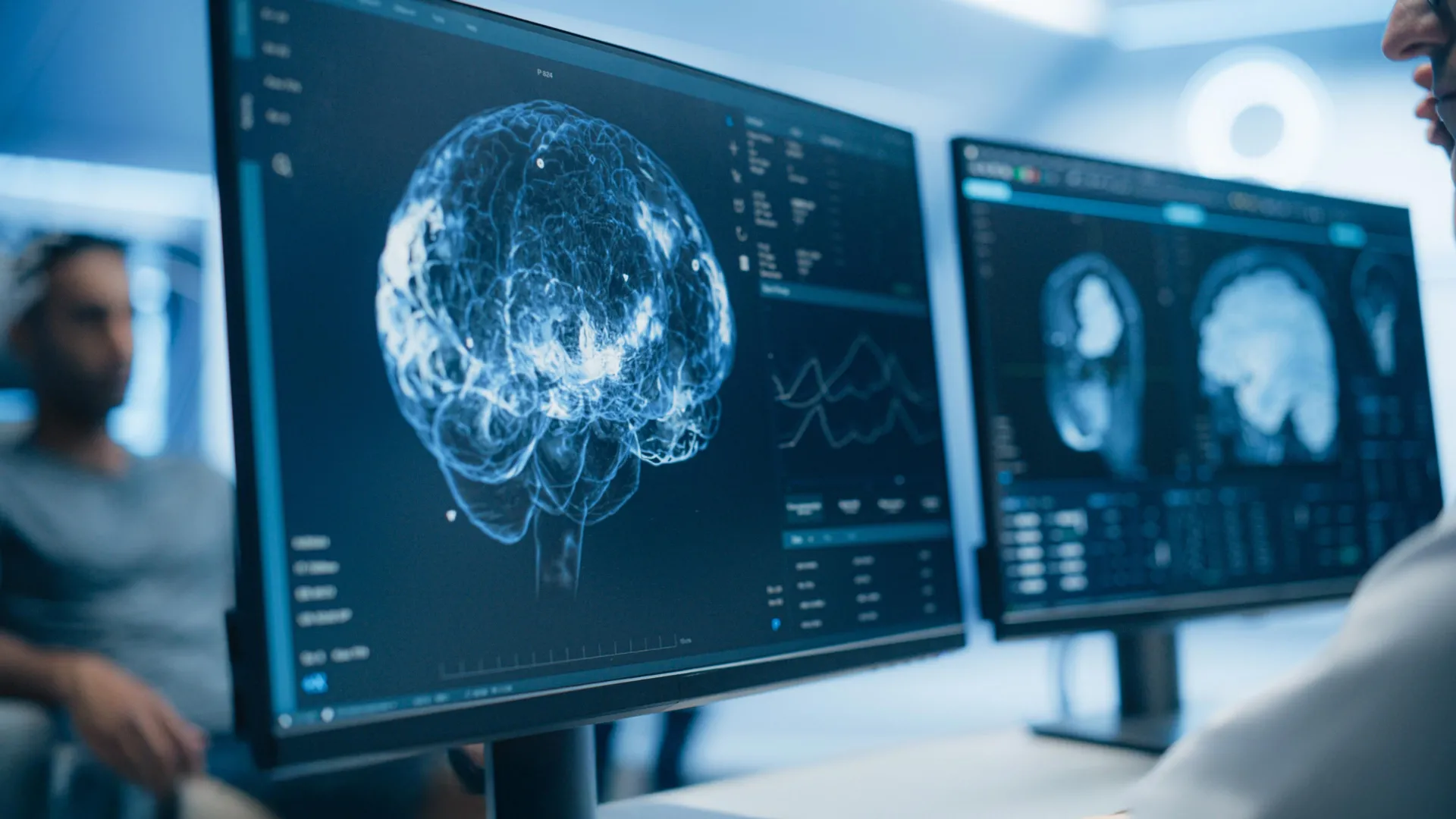
All participants underwent high-resolution Positron Emission Tomography (PET) scans, which are specifically designed to detect the activation of microglial cells—the primary immune cells of the brain. When the brain is inflamed, these cells become activated and express higher levels of certain proteins, such as the translocator protein (TSPO), which the PET scans can identify. In addition to PET imaging, participants received Magnetic Resonance Imaging (MRI) scans to evaluate brain structure and the integrity of white matter, as well as blood tests to measure biomarkers of neuronal damage, such as Neurofilament Light (NfL) and Glial Fibrillary Acidic Protein (GFAP).
Key Findings: The Absence of Chronic Neuroinflammation
The results of the imaging were definitive. When compared to the patients with multiple sclerosis, the long COVID group showed significantly lower levels of inflammatory activity in the brain’s white matter. More strikingly, when the long COVID patients were compared to the healthy control group, the researchers found no statistically significant differences in markers of brain inflammation or neurodegeneration.
"We did not observe evidence of widespread brain inflammation in patients with long COVID when compared to healthy controls," stated Professor Airas, who also serves as a group leader for the InFLAMES Research Flagship. This finding is significant because it suggests that the neurological symptoms of long COVID are not being driven by a persistent "fire" of inflammation in the brain, as many had suspected.
The blood analysis further supported this conclusion. The levels of NfL and GFAP—proteins that spill into the bloodstream when brain cells are damaged or under severe stress—were not elevated in the long COVID group compared to the healthy controls. This indicates that, despite the severity of the symptoms reported by patients, there was no evidence of ongoing structural damage to the neurons or supporting glial cells at the time of the scans.
Chronology of Infection and the Temporal Nature of Inflammation
While the study found no widespread inflammation in the chronic phase, the researchers did uncover a vital clue regarding the timeline of the disease. When analyzing the long COVID cohort, the team noticed a correlation between the time elapsed since the initial infection and the level of inflammatory markers.
Participants who were scanned within 16 months of their initial COVID-19 infection showed slightly higher levels of inflammatory activity in their white matter compared to those who had been ill for a longer period. This suggests a temporal trajectory for the disease: inflammation may indeed be a prominent feature during the acute and sub-acute phases of the illness, but it appears to subside over time.
This chronological insight provides a potential bridge between the new findings and previous neuropathological studies. Earlier autopsies of patients who died during acute, severe COVID-19 infections often showed clear and rampant signs of neuroinflammation. The University of Turku study suggests that for those who survive and transition into long COVID, this inflammatory response may gradually fade, even if the symptoms persist or evolve.
The Role of the Limbic System: Emotion, Stress, and Memory
If inflammation is not the cause, what is driving the symptoms? The study’s most intriguing discovery was found in the brain’s emotional processing centers. The researchers observed that patients who reported the highest levels of anxiety, depression, and a lower quality of life exhibited increased cellular activity in the hippocampus and the amygdala.
These two structures are core components of the limbic system. The hippocampus is essential for memory formation and spatial navigation, while the amygdala is the brain’s primary center for processing emotions, particularly fear and stress. Increased activity in these regions is often associated with a state of heightened physiological stress and emotional dysregulation.
The researchers hypothesize that the symptoms of long COVID may be linked to functional changes in these specific networks. Rather than a generalized inflammatory attack on the brain, the condition may involve a localized "resetting" or over-activation of the brain’s stress-response systems. This could explain why "brain fog"—a symptom often characterized by difficulty concentrating and memory lapses—is so frequently accompanied by emotional symptoms like anxiety.
Shifting the Treatment Paradigm
The implications of these findings for the clinical management of long COVID are profound. For several years, many experimental treatments for long COVID have focused on anti-inflammatory drugs, immunosuppressants, or "blood washing" (apheresis) to remove inflammatory markers. However, if the primary issue in the chronic stage is not inflammation but rather a dysregulation of the limbic system and stress-processing circuits, these treatments may be ineffective for many patients.
The study suggests that a shift in therapeutic focus may be necessary. For patients who have moved past the initial inflammatory phase of the illness, treatments focused on neurological rehabilitation, stress management, and emotional regulation may provide more relief. This does not imply that the condition is "all in the head" in a psychological sense, but rather that the biological mechanism has shifted from an immune-driven problem to a functional neurological one.
Professor Airas emphasized the importance of this shift: "This study highlights the need to continue investigating the complex biological mechanisms underlying long COVID. Understanding these processes is essential for developing targeted treatments."
Broader Impact and Global Health Context
Long COVID remains a major public health crisis, with the World Health Organization (WHO) and the Centers for Disease Control and Prevention (CDC) estimating that between 10% and 30% of people who contract COVID-19 may experience lingering symptoms. The economic impact is equally staggering, as many previously healthy, working-age individuals find themselves unable to return to full-time employment due to cognitive and physical limitations.
The Finnish study adds to a growing body of evidence suggesting that long COVID is a heterogeneous condition—meaning it likely has different causes in different people. While some patients may indeed have persistent viral reservoirs or auto-immune issues, this research identifies a significant subgroup where the primary issue lies in the brain’s functional response to the trauma of the infection.
The research was conducted under the auspices of the InFLAMES Flagship (Innovation Ecosystem based on the Immune System), a joint initiative between the University of Turku and Åbo Akademi University. Supported by the Research Council of Finland, the program is designed to integrate basic immunological research with clinical applications to develop personalized medical treatments. This study represents a key milestone for the initiative, providing a clearer map for future diagnostic and therapeutic interventions.
Future Research Directions
While the study provides critical answers, it also opens new avenues for inquiry. Future research will need to determine why the limbic system remains hyperactive in some patients long after the virus has cleared. Potential areas of study include the role of the autonomic nervous system, the potential for micro-clots to affect localized brain blood flow, and the impact of prolonged social and physical stress on brain plasticity during recovery from a viral illness.
The findings also highlight the importance of the 16-month window identified by the researchers. Future clinical trials for anti-inflammatory therapies may need to target patients specifically within the first year of their symptoms, while trials for neuromodulatory or behavioral therapies might be more appropriate for those in the later stages of the condition.
By moving away from a one-size-fits-all explanation of brain inflammation, the scientific community can begin to develop more precise, individualized care plans for the millions of people still searching for a way to reclaim their health in the wake of the pandemic. The work of Professor Airas and her colleagues serves as a reminder that in the face of a novel disease, the most important step toward a cure is a rigorous, evidence-based understanding of the underlying biology.


Leave a Reply