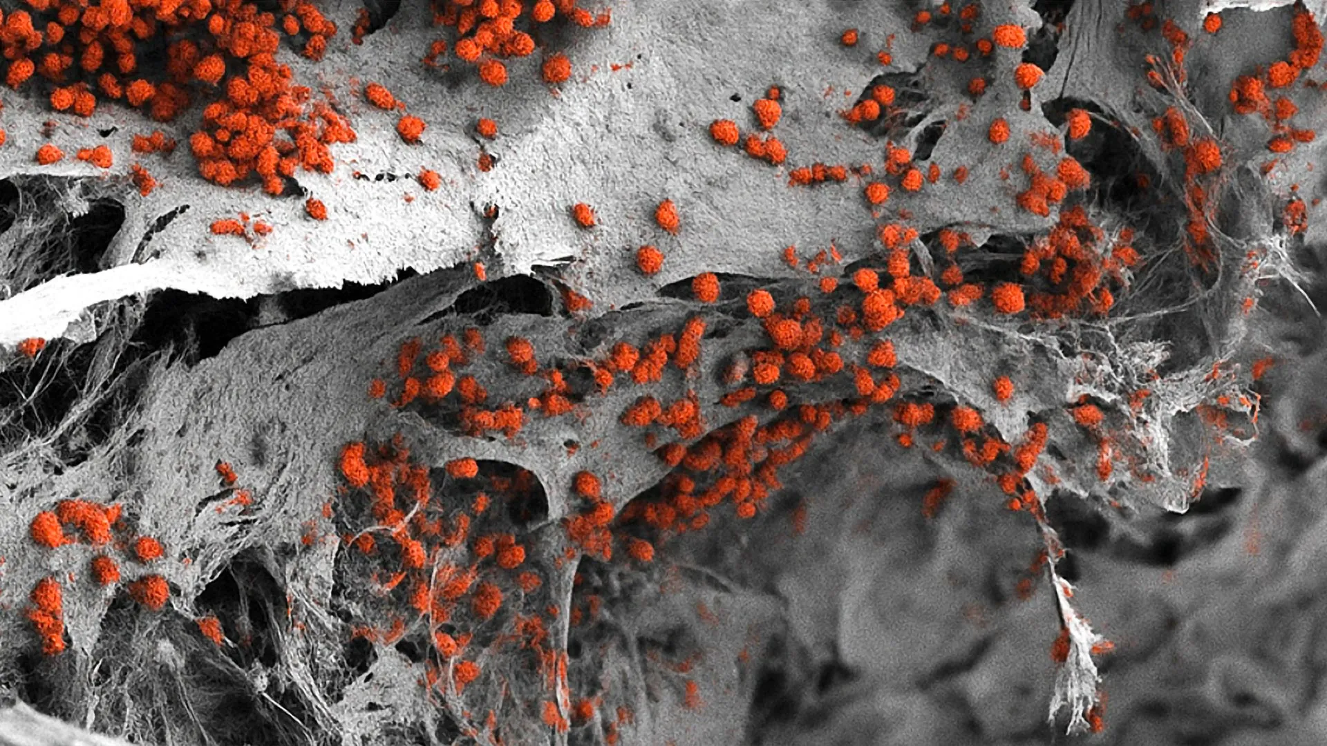
Recent findings published in the prestigious journal Nature have fundamentally shifted our scientific understanding of chronic myeloid leukemia (CML), a rare and aggressive cancer affecting the blood and bone marrow. Conducted by a team of researchers from the Wellcome Sanger Institute alongside international collaborators, the study offers a granular look into the genesis, evolution, and unprecedented multiplication rates of leukemia cells. Utilizing advanced whole-genome sequencing of single blood cells, the scientific team has mapped the precise timeline of how a single genetic aberration can trigger an astonishingly rapid progression toward a clinical diagnosis.
The research illuminates a stark divergence between CML and virtually all other known malignancies, both solid tumors and blood-based cancers. While the conventional paradigm of oncology dictates that cancers develop sluggishly over many decades through the cumulative acquisition of multiple distinct genetic mutations, CML defies this norm. Driven entirely by a single, potent genetic fusion, the disease experiences explosive, exponential growth phases that take clinicians and researchers by surprise. By reconstructing the ancestral lineages of cancer cells, the study provides an unprecedented window into the silent years preceding a formal CML diagnosis.
The Genetic Catalyst: The Philadelphia Chromosome and BCR::ABL1
To comprehend the magnitude of the new findings, it is necessary to examine the foundational biology of chronic myeloid leukemia. CML is fundamentally triggered by a chromosomal translocation—a structural rearrangement in which genetic material is exchanged between two distinct chromosomes. Specifically, a segment of the ABL1 gene located on chromosome 9 is translocated and fused with the BCR gene on chromosome 22.
This abnormal fusion creates a hybrid gene known as BCR::ABL1, which subsequently resides on a shortened chromosome 22, historically designated as the Philadelphia chromosome. The discovery of the Philadelphia chromosome decades ago was a watershed moment in molecular oncology, serving as one of the first direct links between a specific genetic anomaly and the development of cancer. The BCR::ABL1 fusion gene encodes an abnormally active protein—a tyrosine kinase—that continuously signals cells to grow and divide uncontrollably without the normal regulatory constraints that govern healthy cell turnover in the bone marrow.
Despite decades of familiarity with the Philadelphia chromosome and the introduction of targeted therapies that dramatically improved patient survival, significant gaps remained in understanding the natural history of the disease. Prior to this research, the precise rate at which cells carrying the BCR::ABL1 fusion begin to multiply, the exact window of time between the initial genetic event and clinical manifestation, and the subtle variations in evolutionary trajectories among patients remained largely uncharted territory.
Methodology: Tracing Cellular Lineages Through Whole-Genome Sequencing
To pierce the veil of time and observe the historical evolution of CML tumors, the research team adopted a sophisticated genomic approach. The investigators sequenced more than 1,000 whole genomes extracted from single blood cells donated by nine diagnosed CML patients, whose ages ranged from 22 to 81 years old.
By analyzing the unique somatic mutations and genetic signatures present within these individual genomes, the researchers were able to construct detailed phylogenetic trees. Much like genealogical family trees used to trace human ancestry, these cellular phylogenetic trees allowed scientists to trace the lineage of blood cells backward through time. This computational and genetic detective work enabled the team to pinpoint the exact historical moment when the initial BCR::ABL1 gene fusion occurred in a single ancestral blood cell, as well as track how the resulting clone of genetically identical tumor cells expanded over subsequent years.
The resulting timelines proved to be exceptionally illuminating. The phylogenetic reconstruction demonstrated that the BCR::ABL1 fusion typically arises between three and 14 years before a patient receives an official CML diagnosis. This revelation counters any lingering assumptions that the genetic accident occurs merely months before symptoms prompt a clinical evaluation. Instead, the mutation lies dormant or expands quietly beneath the surface for over a decade in many cases, before transitioning into a phase of ferocious growth.
Explosive Growth Rates Defy Conventional Oncology Paradigms
Perhaps the most striking takeaway from the Wellcome Sanger Institute study is the sheer velocity of tumor expansion following the formation of the fusion gene. Once the BCR::ABL1 aberration takes hold, the resulting tumor clones multiply at rates previously undocumented in the broader landscape of oncology.
The data revealed annual growth rates for these clones frequently exceeding 100,000 percent. This blistering pace stands in stark contrast to the development of solid tumors—such as breast, lung, or colorectal cancers—which typically evolve over periods spanning twenty to thirty years through a stepwise accumulation of dozens of driver mutations. Similarly, other hematological malignancies generally progress through gradual clonal evolution.
Furthermore, the study highlights the singular potency of the BCR::ABL1 fusion. While most cancers require a complex symphony of mutations across multiple genes to acquire malignant properties, CML is driven entirely by this singular, highly efficient genetic alteration. The fusion protein acts as a master switch with unprecedented oncogenic strength, single-handedly propelling the rapid accumulation of cancerous white blood cells in the bone marrow and peripheral blood.
Age-Related Dynamics and Treatment Response Implications
In addition to mapping the chronology of the disease, the researchers uncovered critical variations in tumor behavior linked to patient demographics and clinical outcomes. Most notably, age was found to significantly influence the proliferation rate of the cancerous clones. Younger patients exhibited markedly higher multiplication rates of cells harboring the BCR::ABL1 fusion compared to their older counterparts. This biological nuance suggests that the host microenvironment, immune system aging, or physiological differences may modulate the aggressive potential of the leukemia cells.
Crucially, the study bridges the gap between genomic evolution and modern clinical therapeutics. Chronic myeloid leukemia is predominantly managed using targeted therapies known as tyrosine kinase inhibitors (TKIs), which successfully suppress the activity of the BCR::ABL1 protein and have turned a historically fatal diagnosis into a manageable chronic condition for the majority of patients. However, a persistent challenge in hematology is that approximately one in five CML patients fails to achieve an optimal, sustained response to TKI therapy.
The new research sheds light on this clinical hurdle by demonstrating a direct correlation between tumor growth rates and treatment efficacy. Patients whose leukemia cells exhibited faster growth trajectories prior to diagnosis were significantly less likely to respond favorably to standard TKI treatments. By identifying these high-risk growth kinetics at the DNA level, clinicians may eventually be able to stratify patients more accurately upon initial presentation, paving the way for personalized therapeutic adjustments before resistance to standard drugs manifests.
Investigating Asymptomatic Carriers via Large-Scale Cohort Data
To test whether individuals can harbor the BCR::ABL1 fusion gene indefinitely without ever progressing to clinical disease, the researchers expanded their investigation beyond the primary sequencing cohort. They analyzed comprehensive genomic sequencing data and longitudinal electronic health records from more than 200,000 participants enrolled in the expansive, United States-based "All of Us" research program.
The findings from this massive population-level data mining were definitive: virtually all individuals identified as carrying the BCR::ABL1 fusion eventually went on to develop a formal blood disorder or overt leukemia. This indicates that natural immune surveillance or cellular regulatory mechanisms are rarely sufficient to permanently suppress a spontaneously arising BCR::ABL1 clone. Consequently, the silent expansion of these cells without subsequent clinical manifestation is statistically unlikely, reinforcing the notion that the fusion gene possesses an almost inevitable oncogenic destiny once established.
Expert Perspectives and Future Clinical Horizons
The implications of the study have drawn widespread attention from the hematological and oncological communities, offering both a conceptual re-evaluation of cancer biology and practical avenues for future clinical trials.
Dr. Aleksandra Kamizela, co-first author of the study, resident doctor at Lister Hospital in Stevenage, and incoming clinician at Addenbrooke’s Hospital in Cambridge, emphasized the current limitations and future potentials of diagnostic monitoring. "In a clinical setting, healthcare professionals will perform a reverse transcription polymerase chain reaction (RT-PCR) test, a type of blood test, to measure a patient’s response to CML treatment," Dr. Kamizela explained. "However, they are not able to routinely see differences in the genetic cause of CML in patients at the DNA level, which we have been able to highlight in our study. Our findings also provide a rationale to look at the rate of cancer growth more closely in future studies in order to understand if we can use such information in a clinical setting."
Echoing these sentiments, Dr. Jyoti Nangalia, senior author of the study, practicing hematologist at the University of Cambridge, and Group Leader at the Wellcome Sanger Institute, highlighted the exceptional status of CML within the spectrum of human malignancies. "What our study suggests is that chronic myeloid leukemia is an outlier compared to other cancers—both solid tumors and other blood cancers," Dr. Nangalia stated. "We have shown that chronic myeloid leukemia cells undergo incredibly rapid growth within a few years to a decade before diagnosis, whereas for most cancers, the timeline from start to clinical presentation is several decades. This work paves the way to understanding how we might optimize treatment for those patients that currently respond poorly to treatment."
Broader Impact and Concluding Analysis
The publication of this comprehensive genomic analysis marks a pivotal milestone in the ongoing effort to outsmart hematological malignancies. By detailing the subterranean timeline of CML development—revealing that the cancer quietly establishes its roots over three to fourteen years before erupting in explosive, hyper-proliferative growth phases—the Wellcome Sanger Institute team has dismantled long-held assumptions regarding the uniformity of cancer evolution.
While the study opens exciting pathways for optimizing therapeutic strategies for the minority of patients who struggle with standard TKI treatments, the authors caution that further validation through larger, prospective patient cohorts will be essential before these genomic insights can be fully integrated into routine clinical guidelines. Nevertheless, by bridging the gap between high-resolution single-cell genomics and real-world oncology, this research provides a robust foundation for more precise, individualized management of chronic myeloid leukemia, ultimately bringing medical science one step closer to neutralizing one of blood cancer’s most deceptive outliers.