
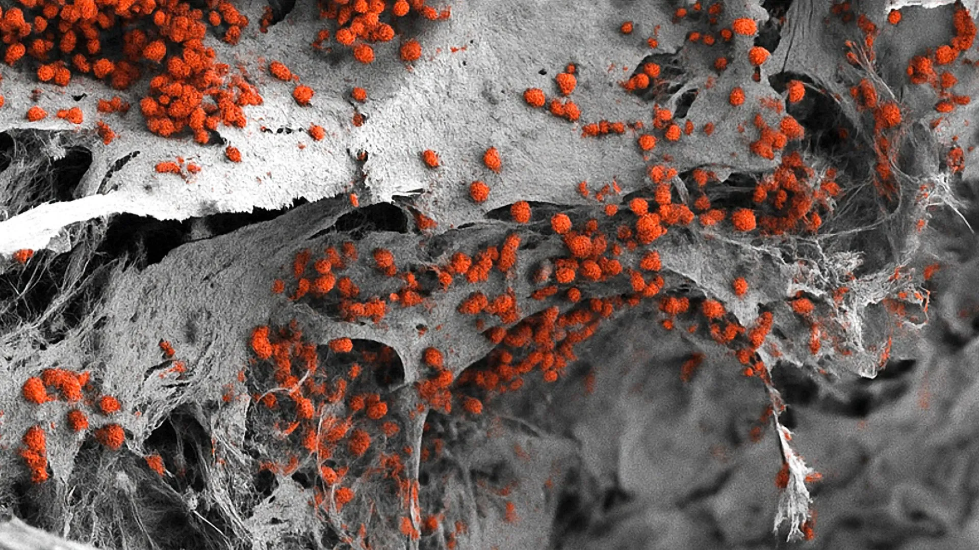
The landscape of biomedical research and hematological studies has reached a significant milestone following a groundbreaking achievement by a collaborative team of scientists at the University of Basel and the University Hospital Basel. For the first time in the history of cellular biology, researchers have successfully engineered a fully functional, highly complex human bone marrow system derived exclusively from human cells. This sophisticated three-dimensional biological model mirrors the intricate architecture of the human body’s primary blood-manufacturing organ, incorporating bone cells, specialized neural networks, vascular pathways, and a diverse array of immune components. Published in the esteemed scientific journal Cell Stem Cell, this development addresses a long-standing limitation in medical science: the inability to accurately replicate the human bone marrow microenvironment in vitro without relying on animal models or overly simplistic cellular assays.
Often referred to as the body’s blood factory, the bone marrow usually operates in a state of quiet equilibrium, hidden deep within the skeletal structure. It generates billions of red blood cells, white blood cells, and platelets daily to sustain life, yet it remains largely invisible to the conscious mind until its delicate processes begin to fail. Disorders such as leukemia, multiple myeloma, and various forms of lymphoma disrupt this equilibrium, transforming the marrow into a site of pathological dysfunction. Historically, investigating the root causes of these hematological malignancies and understanding how resistance to chemotherapy develops has required invasive human biopsies or extensive animal testing, primarily utilizing murine models. While mice have historically provided invaluable insights into mammalian hematopoiesis, significant physiological differences between rodents and humans have frequently hindered the translation of laboratory discoveries into successful clinical therapies. The newly unveiled human cell model bridges this translational gap, offering a physiologically relevant, human-specific testing ground that promises to accelerate the discovery of life-saving interventions while aligning with the global scientific mandate to reduce, refine, and replace animal experimentation.
Unraveling the Complexities of the Endosteal Niche
At the heart of this scientific breakthrough is the accurate replication of specific microenvironments within the bone marrow, known scientifically as niches. The bone marrow is not a uniform soup of cells; rather, it is a highly organized organ divided into distinct zones that regulate stem cell behavior, differentiation, and maintenance. Among these, the endosteal niche—situated immediately adjacent to the inner surface of the bone—plays a critically important and notoriously complex role. This specialized zone is a bustling cellular metropolis consisting of osteoblasts (bone-forming cells), vascular endothelial cells, neural fibers, and various immune cells working in concert.
Crucially, the endosteal niche is deeply implicated in the pathogenesis of blood cancers. Scientific literature has repeatedly demonstrated that cancer cells often migrate to this specific microenvironment, where they find sanctuary from systemic chemotherapy and radiation treatments. This protective sanctuary allows residual malignant cells to survive, leading to disease relapse and treatment failure. Until now, recreating this multifaceted niche in a laboratory setting had eluded researchers. Previous in vitro models could capture isolated elements—such as individual cell types or simplified two-dimensional layers—but failed to encapsulate the structural and cellular crosstalk that defines the native human endosteal environment.
The research team, spearheaded by Professor Ivan Martin and Dr. Andrés García García from the Department of Biomedicine, overcame this hurdle by adopting a multi-step bioengineering strategy. Their methodology began with the construction of an artificial bone scaffold fabricated from hydroxyapatite, a naturally occurring calcium phosphate mineral that forms the primary structural matrix of human bones and teeth. This scaffold provided the necessary mechanical rigidity and biochemical cues required to support bone tissue formation and cellular attachment.
Reprogramming Cellular Destinies through Stem Cell Technology
To populate this mineral framework, the researchers turned to advanced molecular biology techniques, specifically utilizing human pluripotent stem cells. These remarkable cells possess the rare ability to differentiate into virtually any cell type found in the human body, provided they receive the correct biochemical instructions and environmental cues. By taking somatic human cells—such as skin or blood cells—and reprogramming them into induced pluripotent stem cells (iPSCs), the scientific team unlocked an unlimited, ethically sound cellular resource.
The researchers meticulously introduced these pluripotent stem cells into the porous hydroxyapatite scaffold. Through carefully controlled, stepwise biochemical stimulation, the stem cells were guided to differentiate into the diverse lineage of cells native to the bone marrow. Over a period of weeks, these cells self-organized within the three-dimensional matrix, establishing intricate networks of blood vessel precursors, neural elements, and bone-producing cells.
Unlike previous micro-models that resembled microscopic specks, this newly engineered system boasts macro-scale dimensions. Measuring an impressive eight millimeters in diameter and four millimeters in thickness, the construct is significantly larger and more robust than its predecessors. Most importantly, histological and functional analyses confirmed that the internal architecture of the engineered tissue closely mimics the native human endosteal niche. Within this artificial sanctuary, the researchers successfully maintained human blood cell formation—hematopoiesis—for a period of several weeks, proving that the system is not merely structurally accurate, but metabolically and functionally active.
Chronology of the Breakthrough and Collaborative Effort
The realization of this project represents the culmination of years of methodical research in tissue engineering, stem cell biology, and biomaterials science at the University of Basel. The foundational work involving hydroxyapatite scaffolds and stem cell differentiation pathways began nearly a decade ago, building upon incremental advances in regenerative medicine. As biofabrication techniques matured, the team steadily increased the complexity of their cellular constructs, moving from simple two-dimensional co-cultures to primitive three-dimensional models, and finally achieving the multi-lineage integration seen in the current Cell Stem Cell publication.
The collaborative framework between the University of Basel and the University Hospital Basel was instrumental in bridging basic scientific discovery with clinical relevance. By drawing upon clinical insights regarding blood cancer progression and treatment resistance, the bioengineers were able to tailor their model to address specific diagnostic and therapeutic bottlenecks faced by hematologists and oncologists daily. The peer-review process, which culminated in the acceptance of the study by leading international reviewers, further validates the methodological rigor and reproducibility of the Basel team’s findings.
Implications for Animal Testing and Regulatory Shifts
A prominent driving force behind the development of this human cell-based bone marrow model is the growing ethical and scientific pressure to minimize animal research. For decades, preclinical drug development has depended heavily on murine models to evaluate toxicity, pharmacokinetics, and therapeutic efficacy. However, species-specific differences in immune system responses, bone marrow architecture, and metabolic pathways frequently result in high attrition rates when animal data fails to translate successfully to human clinical trials.
Professor Ivan Martin emphasizes the transformative potential of the new platform in this context. While acknowledging the historical debt that modern medicine owes to mouse studies in unraveling the fundamental principles of hematopoiesis, Martin points out that the Basel model brings researchers substantially closer to the authentic biology of the human organism. By providing a human-centric testing environment, the system offers a viable, scientifically superior complement to numerous animal experiments.
This development resonates strongly with the broader institutional and international initiatives aimed at implementing the Three Rs principle: Replacement, Reduction, and Refinement of animal use in scientific research. Regulatory bodies across Europe, North America, and other jurisdictions are increasingly receptive to advanced alternative methods—such as organ-on-a-chip technologies, complex 3D tissue models, and human cell assays—provided they can demonstrate robust predictive validity. While complete replacement of animal models remains a long-term aspiration rather than an immediate reality, tools like the Basel bone marrow model represent monumental strides toward achieving that goal.
Challenges in Scalability and High-Throughput Drug Screening
Despite its impressive biological fidelity, the current iteration of the bone marrow model presents certain technical challenges that must be addressed before it can be seamlessly integrated into commercial pharmaceutical pipelines. One notable hurdle relates to its physical dimensions. Measuring nearly a centimeter across, the model is exceptionally well-suited for detailed cellular analysis, long-term culture maintenance, and the observation of complex tissue dynamics. However, these very dimensions render it less than ideal for high-throughput screening applications.
Dr. Andrés García García highlights this operational constraint, noting that modern drug discovery workflows often require the simultaneous testing of hundreds of distinct compounds, drug combinations, or concentration gradients within tight timeframes. To adapt the current platform for such high-throughput screening, the research team must successfully miniaturize the system without compromising its cellular diversity or structural complexity. Scaling down the architecture while maintaining the intricate crosstalk between the endosteal niche components represents the next major engineering frontier for the Basel laboratory.
Pathways to Personalized Medicine and Oncological Innovation
Looking toward the horizon, the most profound potential of this technology lies in the realm of personalized medicine. Treating hematological malignancies remains a formidable challenge due to the significant genetic and phenotypic heterogeneity observed among patients diagnosed with the same broad category of blood cancer. What proves to be a curative regimen for one individual may fail entirely in another, often forcing oncologists to rely on trial-and-error treatment strategies.
To overcome this limitation, the research team envisions a future where clinicians could utilize patient-specific cells—harvested safely through routine biopsies or blood draws—to construct customized, individual bone marrow models in the laboratory. By introducing a patient’s own malignant cells into their personalized artificial niche, medical professionals could directly test a panel of chemotherapeutic agents, targeted therapies, or immunotherapies against the live tissue model. This empirical testing would allow oncologists to identify the most efficacious treatment strategy tailored specifically to the unique biological profile of that patient’s disease before administering a single dose in the clinic.
While realizing this vision of personalized oncology will require substantial technological refinement, workflow standardization, and rigorous clinical validation, the foundational proof-of-concept has now been firmly established. By successfully uniting advanced biomaterials with pluripotent stem cell technology, the University of Basel team has opened a promising new chapter in hematological research. As this technology matures and scales, it holds the genuine potential to transform how blood disorders are understood, how pharmaceutical compounds are evaluated, and how personalized cancer therapies are administered in the modern medical era.