In a landmark study that bridges the fields of oncology and neurology, researchers at Boston Children’s Hospital have identified a startling genetic link between the development of Alzheimer’s disease and the mutations typically associated with blood cancers. The research, published in the journal Cell, suggests that microglia—the specialized immune cells of the central nervous system—accumulate somatic mutations in specific cancer-driving genes as they age. However, instead of forming malignant tumors, these mutated cells appear to exacerbate the neurodegenerative processes that lead to Alzheimer’s disease.
The study, led by Christopher Walsh, MD, PhD, Chief of the Division of Genetics and Genomics at Boston Children’s Hospital and an Investigator of the Howard Hughes Medical Institute, fundamentally alters the scientific understanding of how Alzheimer’s progresses. By identifying that these mutations are also present in the bloodstream of patients, the research opens a new frontier for non-invasive diagnostic testing and the potential repurposing of existing oncology drugs to treat dementia.
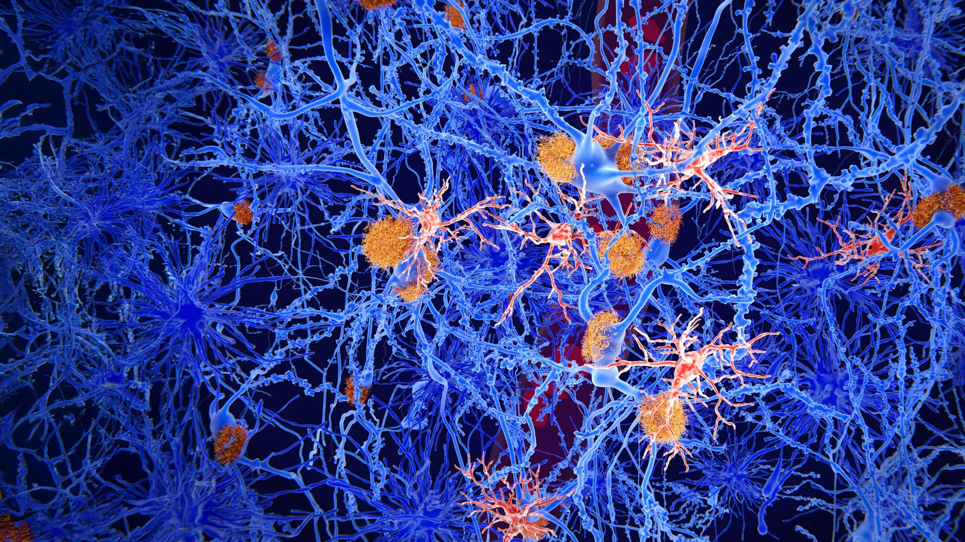
The Role of Microglia and Somatic Mosaicism
Microglia have long been recognized as the "cleanup crew" of the brain. Under normal physiological conditions, these cells are responsible for maintaining neural health by scavenging for damaged neurons, clearing cellular debris, and neutralizing infectious agents. Unlike other immune cells that circulate throughout the body, microglia were historically believed to be a stable population that remains sequestered within the brain, protected by the blood-brain barrier (BBB).
As humans age, their cells undergo a process known as somatic mosaicism. While germline mutations are inherited from parents and present in every cell, somatic mutations are acquired throughout a person’s lifetime due to environmental factors, errors in DNA replication, or aging. These mutations are often localized to specific tissues. The research team at Boston Children’s Hospital set out to determine if these accumulated mutations in the brain played a role in age-related cognitive decline.
"We find that to some extent, Alzheimer’s disease is a little like cancer—driven by the same mutations that drive blood cancers like lymphoma and leukemia," stated Dr. Walsh. This conceptual shift suggests that the neuroinflammation seen in Alzheimer’s may not just be a reaction to protein plaques, but a result of a "clonal expansion" of mutant immune cells that have become dysfunctional or overly aggressive.
Methodology: Analyzing the Genetic Landscape of the Aging Brain
To reach these conclusions, the research team conducted an exhaustive genetic analysis of brain tissue samples. The study involved 190 individuals diagnosed with Alzheimer’s disease and a control group of 121 individuals with healthy brains. The researchers focused their investigation on 149 known cancer-driving genes, looking for "single-letter" DNA changes—point mutations that can alter the function of a protein.
The results revealed a significant disparity between the two groups. The Alzheimer’s samples contained a much higher frequency of somatic mutations than the healthy tissue. Most notably, these mutations were not randomly distributed; they repeatedly appeared in a specific set of five cancer-driving genes. This pattern suggested that cells harboring these specific mutations were being "selected" for, allowing them to multiply more rapidly than their non-mutated counterparts within the brain environment.
The Unexpected Link Between Blood and Brain
The most surprising discovery occurred when the researchers examined the origins of these mutated microglia. Because the mutations identified were identical to those found in blood cancers like leukemia, the team decided to cross-reference the brain findings with blood samples from the same Alzheimer’s patients.
Conventional medical wisdom suggested that the blood and brain immune systems were largely separate. However, the researchers found the exact same cancer-associated mutations in the blood cells of the Alzheimer’s patients. This finding led to a new hypothesis regarding the pathogenesis of the disease: the mutant cells were not originating in the brain but were migrating there from the bone marrow and bloodstream.
"It was actually a really unexpected finding that suggests a totally new mechanism for Alzheimer’s disease pathogenesis," said August Yue Huang, PhD, a co-author of the study and professor at Harvard Medical School. "The findings mean that the blood’s immune cells with cancer mutations are likely getting into the brain and contributing to disease."
Chronology of Pathogenesis: How Mutant Cells Infiltrate the CNS
The researchers propose a multi-stage timeline for how these mutant cells contribute to Alzheimer’s:
- Systemic Mutation: As an individual ages, somatic mutations occur in hematopoietic stem cells (the precursors to blood cells) in the bone marrow. This is a known phenomenon called Clonal Hematopoiesis of Indeterminate Potential (CHIP).
- Barrier Breakdown: Under normal circumstances, the blood-brain barrier prevents these mutant blood cells from entering the central nervous system. However, aging, chronic hypertension, or localized brain injuries can weaken this barrier.
- Infiltration and Transformation: Once the barrier is compromised, these mutant immune cells enter the brain. Once inside, they transform into cells that look and act like microglia.
- Clonal Expansion: In an Alzheimer’s brain, the presence of amyloid-beta plaques and tau tangles creates a high-stress, inflammatory environment. The mutant cells, which possess a survival advantage due to their cancer-driving genetics, begin to proliferate more rapidly than healthy microglia.
- Neurotoxic Environment: These mutated, "cancer-like" microglia are likely more inflammatory and less efficient at clearing debris than healthy cells. Their presence creates a toxic environment that leads to the death of nearby neurons, accelerating cognitive decline.
Supporting Data: Independence from APOE4
One of the most significant aspects of this research is its relationship to known genetic risk factors. For decades, the APOE4 allele has been the primary genetic marker for Alzheimer’s risk. However, many people with APOE4 never develop the disease, while many without it do.
In a follow-up study published as a preprint on bioRxiv, Dr. Huang and Dr. Alice Eunjung Lee found that the presence of these cancer-driver mutations in the blood increased the risk of Alzheimer’s disease independently of the APOE4 status. This suggests that somatic mutations in the blood may be a "second hit" or a separate pathway to the disease entirely, explaining why some individuals with low hereditary risk still succumb to the condition.
The data indicates that these mutations could serve as a missing piece of the puzzle in understanding the "sporadic" form of Alzheimer’s, which accounts for the vast majority of cases and has historically been difficult to predict through traditional genetics.
Implications for Diagnosis and Therapeutic Intervention
The discovery that Alzheimer’s-linked mutations are detectable in the blood has profound implications for early diagnosis. Currently, diagnosing Alzheimer’s often requires expensive PET scans or invasive lumbar punctures to detect amyloid and tau levels.
"Because it’s hard to access brain tissue in a living patient, genetic screens using blood samples could be developed to test whether a person carries these mutations, and has an increased risk of developing Alzheimer’s disease," explained Dr. Alice Eunjung Lee. Such a "liquid biopsy" for Alzheimer’s could allow for screening decades before symptoms appear, providing a window for preventative intervention.
Furthermore, the link to cancer genetics provides a ready-made library of potential treatments. The pharmaceutical industry has already developed dozens of FDA-approved drugs designed to target the specific pathways activated by these cancer-driving mutations.
"This is helpful because we have a lot of drugs to fight cancer and some of them might be useful therapeutically for Alzheimer’s disease," Walsh noted. For instance, drugs that inhibit the proliferation of mutant white blood cells in leukemia might be repurposed to slow the expansion of toxic microglia in the brain, effectively "treating" the neuroinflammation by treating it like a slow-growing, localized immune expansion.
Collaborative Efforts and Future Outlook
The research was a massive collaborative effort involving Boston Children’s Hospital, Harvard Medical School, the Broad Institute of MIT and Harvard, and the Icahn School of Medicine at Mount Sinai. The study was supported by significant funding from the Howard Hughes Medical Institute and the National Institute on Aging.
The work is also part of the Somatic Mosaicism Across Human Tissues (SMaHT) consortium, an NIH Common Fund initiative aimed at mapping how somatic mutations contribute to human health and disease across different organs.
While the findings are transformative, researchers caution that more work is needed to determine exactly which cancer drugs might be effective and at what stage of the disease they should be administered. However, the shift in perspective—viewing Alzheimer’s not just as a protein-folding problem but as a condition influenced by the "oncological" behavior of the immune system—marks a new era in dementia research.
As the global population ages and the prevalence of Alzheimer’s is expected to rise, the ability to identify high-risk individuals through a simple blood test and treat them with established oncology protocols could represent one of the most significant breakthroughs in the history of geriatric medicine. The study serves as a potent reminder that the boundaries between different fields of medicine, such as oncology and neurology, are often thinner than previously imagined.