In a landmark achievement for regenerative medicine and developmental biology, an international consortium of researchers has published the first comprehensive "blueprint" of the human skeletal system’s earliest stages. This exhaustive map, part of the monumental Human Cell Atlas (HCA) initiative, provides an unprecedented look at how the human frame is constructed in the womb, offering transformative insights into the origins of arthritis, rare bone deformities, and the potential risks of pharmaceutical interference during pregnancy. Published on November 20 in the journal Nature, the study represents a significant leap in our understanding of human biology, detailing the cellular pathways and genetic triggers that govern bone formation from the fifth to the eleventh week post-conception.
The research, led by the Wellcome Sanger Institute in collaboration with several global partners, utilized cutting-edge single-cell genomics and spatial transcriptomics to chart the odyssey of skeletal development. By documenting the precise location and activity of every cell type involved in the process, the team has created a spatial and temporal resource that is now freely available to the global scientific community. This atlas is not merely a catalog of cells; it is a functional guide that explains why certain joints fail in old age and how genetic mutations can cause the skulls of newborns to fuse prematurely, a condition that can have devastating neurological consequences.
The Chronology of Human Skeletal Construction
The development of the human skeleton is a highly coordinated biological symphony that begins shortly after conception. The researchers focused their efforts on the first trimester—specifically the window between 5 and 11 weeks—which is the critical period of organogenesis where the basic structures of the body take shape. During this time, the skeleton undergoes a process known as endochondral ossification for the majority of the body, and intramembranous ossification for specific areas like the skull.
According to the study’s findings, the vast majority of the human skeleton begins not as bone, but as a flexible scaffold of cartilage. This cartilage template acts as a placeholder, providing the necessary structure while bone cells (osteoblasts) gradually move in to replace the softer tissue. The researchers mapped this transition with granular detail, identifying the "transition zones" where cartilage cells mature and eventually signal for bone mineralization.
However, the team discovered a notable exception to this rule: the calvarium, or the upper part of the skull. Unlike the long bones of the arms or the vertebrae of the spine, the top of the skull does not utilize a cartilage scaffold. Instead, specialized early bone cells form directly from mesenchymal tissue. This distinction is critical for understanding how the human head evolves to accommodate the rapidly expanding brain of an infant.
Decoding Craniosynostosis and Skull Development
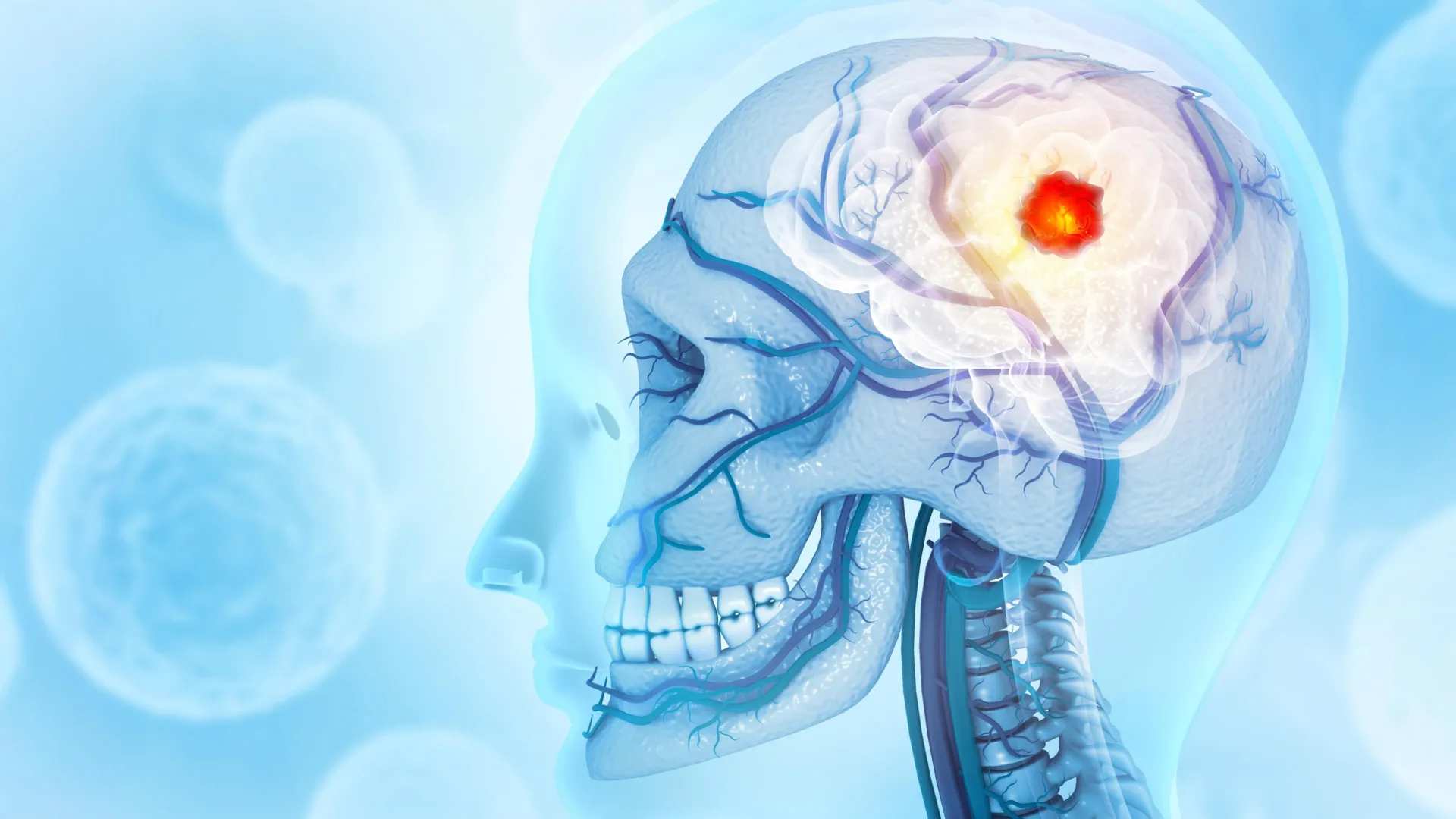
One of the most immediate clinical applications of the new atlas involves craniosynostosis, a condition where the "soft spots" (fontanelles) and sutures of an infant’s skull fuse too early. In a healthy development cycle, these gaps remain open for one to two years after birth to allow for brain growth. If they close prematurely, the resulting pressure can lead to permanent brain damage, vision loss, and hearing impairment.
While the medical community has long known that craniosynostosis is linked to specific genetic mutations, the exact cells targeted by these mutations remained a mystery. The new HCA data has finally identified the specific populations of early bone cells in the calvarium that are disrupted by these genetic errors. By mapping these interactions, researchers have opened the door to new diagnostic tools that could identify at-risk pregnancies and potentially lead to non-surgical interventions in the future.
In the United Kingdom, where the study was centered, craniosynostosis is currently treated through complex reconstructive surgery. The ability to target the cellular mechanisms behind the condition could eventually reduce the need for such invasive procedures.
The Genetic Link Between Fetal Development and Adult Arthritis
Perhaps the most surprising revelation from the skeletal atlas is the connection between the first trimester of pregnancy and the development of osteoarthritis (OA) decades later. Osteoarthritis is the most prevalent form of arthritis worldwide, characterized by the breakdown of protective cartilage in the joints. Because adult humans lack the ability to naturally regenerate damaged cartilage, severe cases often necessitate total joint replacement.
The research team cross-referenced their developmental data with known genetic risk factors for osteoarthritis. They found a striking divergence in how the disease manifests in different joints:
- Hip Arthritis: Genetic variants associated with a higher risk of hip OA were found to be active in the very early stages of bone cell development. This suggests that the structural integrity of the hip joint may be "programmed" during the first trimester, with subtle developmental variations predisposing individuals to joint failure in later life.
- Knee Arthritis: In contrast, genetic variants linked to knee OA were found to be active in cartilage formation and repair pathways. This implies that knee arthritis is more closely tied to the body’s inability to maintain and fix the cartilage scaffold throughout an individual’s lifespan.
"Having this ‘blueprint’ of bone formation can help us develop effective ways to grow bone and cartilage cells in a dish, which has enormous therapeutic potential," noted Dr. Ken To, co-first author from the Wellcome Sanger Institute. This insight could revolutionize the field of regenerative orthopedics, allowing scientists to engineer "young" cartilage that mimics the resilient properties of fetal tissue.
Pharmacological Safety and the Developing Skeleton
Beyond disease pathology, the skeletal atlas serves as a vital safety manual for modern medicine. The researchers used the data to evaluate the impact of 65 clinically-approved drugs that are currently flagged as potentially hazardous or not recommended during pregnancy. By overlaying the chemical pathways of these drugs with the gene networks active in the developing skeleton, the team could pinpoint exactly where and how these medications might disrupt bone growth.
This aspect of the study is particularly relevant for expectant mothers who may require treatment for chronic conditions. Current pharmaceutical guidelines are often based on animal studies or retrospective human data, which can be imprecise. The HCA skeletal atlas provides a direct, human-specific model to test the safety of therapeutics, potentially leading to more nuanced prescribing guidelines that protect the developing fetus while ensuring the health of the mother.
The Human Cell Atlas: A Global Scientific Milestone
The skeletal blueprint is not an isolated discovery; it is part of a massive coordinated release of over 40 publications in Nature Portfolio journals, all contributing to the Human Cell Atlas. The HCA is an international collaborative effort to map every cell type in the human body—from the brain to the immune system—to create a "Google Maps" of human biology.
Professor Sarah Teichmann, co-founder of the HCA and senior author of the study, emphasized the collaborative nature of the project. "Our unique freely available skeletal atlas sheds new light on cartilage, bone, and joint development in the first trimester, detailing the cells and pathways involved together for the first time," she stated. "This detailed atlas is coordinated with other studies which brings the entire Human Cell Atlas initiative one step closer to fully understanding what happens in the human body across development, health, and disease."
The release of these 40+ papers marks a milestone leap in the field of genomics. By combining spatial transcriptomics—which shows where genes are active in a tissue—with single-cell sequencing, researchers can now see the "architectural plans" of life in high definition.
Analysis of Broader Implications and Future Research
The implications of this research extend far into the future of healthcare. For the pharmaceutical industry, the atlas provides a new platform for drug discovery, allowing for the screening of compounds against specific human fetal cell types without the ethical and logistical hurdles of traditional clinical trials. For the field of evolutionary biology, it provides clues as to how the human skeleton evolved its unique characteristics, such as the upright posture and the large cranium.
Furthermore, the study highlights the importance of the first trimester as a foundation for lifelong health. The finding that adult-onset diseases like arthritis have roots in fetal development shifts the paradigm of "age-related" conditions, suggesting that early-life interventions or monitoring could prevent chronic disability in the elderly.
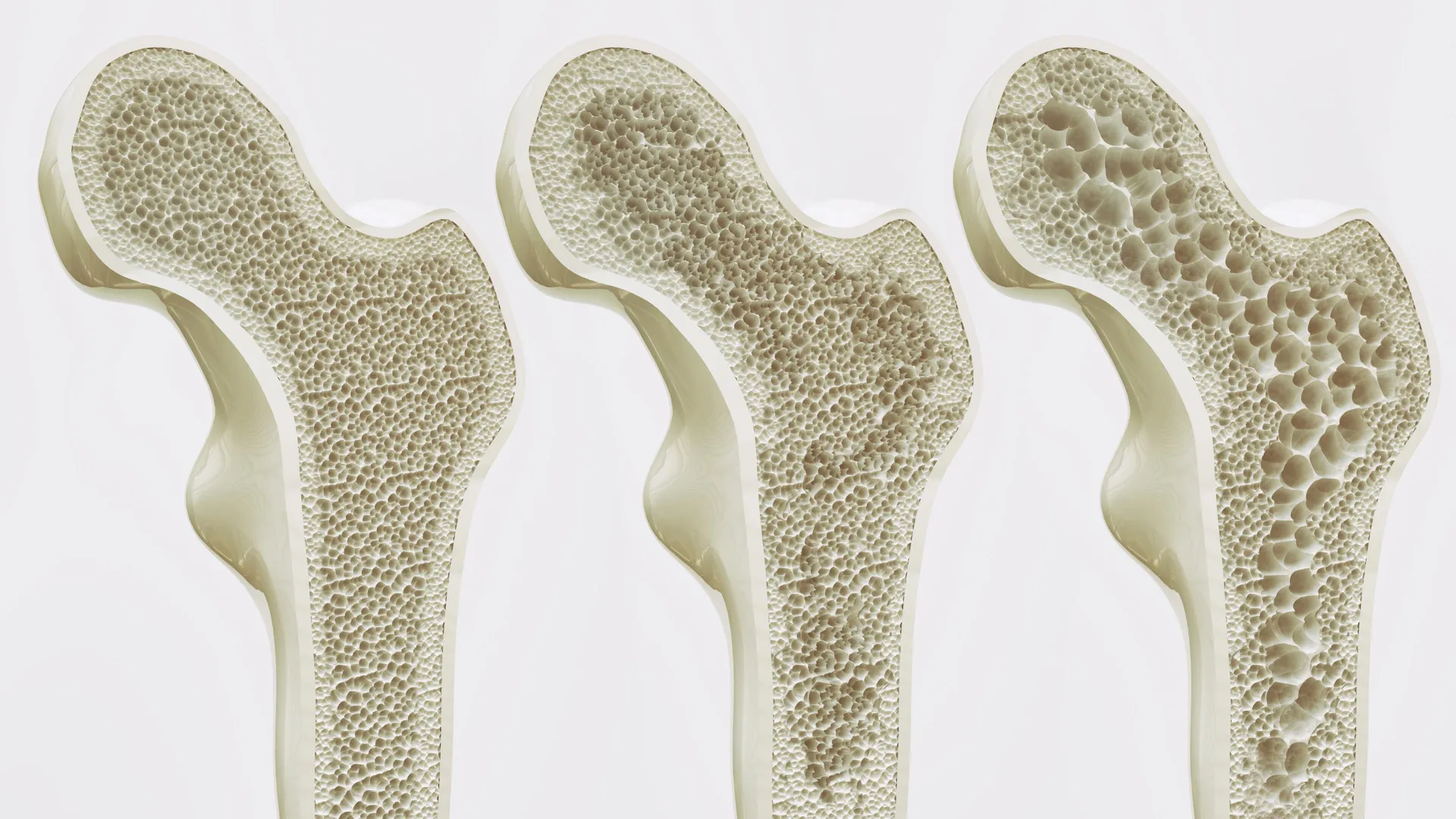
Dr. Jan Patrick Pett, co-first author, highlighted the computational power behind the project: "Our multi-layered, time- and space-resolved atlas enabled novel computational analyses, which we used to create an integrated view of how developmental processes are regulated." This integrated view is expected to fuel a decade of follow-up studies, as researchers worldwide begin to mine the data for clues to other musculoskeletal conditions, such as scoliosis, osteoporosis, and rare bone cancers.
As the scientific community begins to digest this wealth of information, the message is clear: the journey to understanding the human body does not begin at birth, but in the silent, rapid cellular divisions of the first few weeks of life. By mapping those origins, the Human Cell Atlas has provided a map that will guide medical science for generations to come.