In a landmark achievement for regenerative medicine and hematology, researchers from the University of Basel and University Hospital Basel have successfully engineered a fully functional, three-dimensional model of human bone marrow using exclusively human cells. This breakthrough, recently detailed in the journal Cell Stem Cell, represents the first time scientists have been able to replicate the intricate "blood factory" of the human body in a laboratory setting without relying on animal-derived components or simplified two-dimensional cultures. Led by Professor Ivan Martin and Dr. Andrés García García from the Department of Biomedicine, the team has provided a sophisticated platform that could revolutionize how we study blood-related diseases, test new pharmacological compounds, and eventually deliver personalized therapies for cancer patients.
The human bone marrow is one of the most complex tissues in the body, serving as the primary site for hematopoiesis—the continuous process of blood cell production. It is a highly specialized environment where bone cells, nerves, blood vessels, and various immune cells interact within a precise structural framework. Despite its critical role in sustaining life, the bone marrow often remains a "black box" in clinical research, only becoming a primary focus when its delicate balance is disrupted by malignancies such as leukemia, multiple myeloma, or myelodysplastic syndromes. For decades, the scientific community has struggled to recreate this environment, often forced to choose between animal models, which do not always accurately reflect human physiology, and basic cell cultures that lack the three-dimensional architecture necessary for realistic biological signaling.
The Evolution of Bone Marrow Modeling: A Chronological Perspective
The journey toward this breakthrough began decades ago with the realization that the bone marrow environment, or "niche," is as important as the stem cells themselves. In the mid-20th century, early hematology research focused on identifying the hematopoietic stem cell (HSC), the progenitor of all blood cells. By the 1970s and 1980s, researchers began to understand that HSCs require specific signals from their surroundings to decide whether to self-renew or differentiate into oxygen-carrying red cells, infection-fighting white cells, or clot-forming platelets.
Throughout the 1990s and early 2000s, the gold standard for studying these processes was the mouse model. While murine studies provided foundational knowledge, they often failed to translate into successful human clinical trials. A significant turning point occurred in 2006 with the discovery of induced pluripotent stem cells (iPSCs), which allowed scientists to "reprogram" adult human cells back into a primitive state capable of becoming any cell type. This technology provided the raw materials for the Basel team’s current success. Over the last ten years, tissue engineering has moved from simple cell clusters to complex "organ-on-a-chip" systems, but replicating the dense, mineralized environment of the bone surface—the endosteal niche—remained a significant hurdle until now.
Engineering the Endosteal Niche: Technical Innovations and Data
The Basel researchers focused their efforts on the endosteal niche, a specific microenvironment located near the inner surface of the bone. This area is of particular interest to oncologists because it is believed to be a "sanctuary" where cancer cells can hide from chemotherapy, leading to disease relapse. To replicate this, the team utilized a multi-step bioengineering approach.
The foundation of the model is an artificial scaffold made of hydroxyapatite. This mineral, a naturally occurring form of calcium apatite, constitutes the bulk of human bone and tooth enamel. By using hydroxyapatite, the researchers provided the cells with the physical and chemical cues they would encounter in a living body. The team then introduced human iPSCs into this scaffold. Through a carefully orchestrated series of molecular signals and growth factors, they guided these stem cells to differentiate into the diverse array of cell types found in the marrow, including osteoblasts (bone-forming cells), endothelial cells (which form blood vessels), and mesenchymal stromal cells.
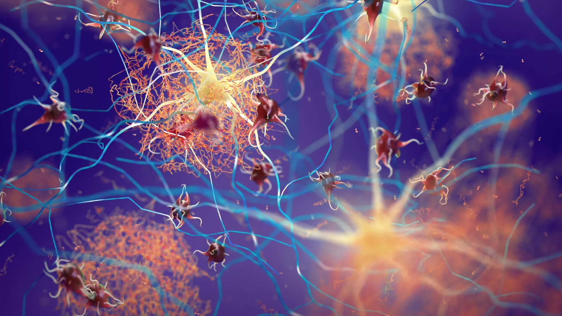
The resulting structure is a feat of micro-engineering. Measuring approximately eight millimeters in diameter and four millimeters in thickness, the model is significantly larger and more complex than previous iterations. Data analysis of the engineered tissue confirmed that it contains a functional network of blood vessels and neural-like structures, mirroring the vascular and nervous integration found in native bone marrow. Most importantly, the model successfully maintained the production and maturation of human blood cells for several weeks in a laboratory incubator, a duration that allows for extended observation and testing.
Addressing the Limitations of Animal Experimentation
One of the primary drivers of this research is the global movement to reduce, refine, and replace (the 3Rs) animal testing in scientific research. While mouse models have been instrumental in biological discovery, the physiological discrepancies between species are substantial. For instance, the signaling molecules (cytokines) that regulate blood production in mice do not always cross-react with human cells, and the architecture of mouse bone is significantly different in scale and density.
Professor Ivan Martin emphasized that while mouse studies have provided a roadmap, the new human-cell model brings researchers much closer to the specific biology of the human organism. By using 100% human cells, the researchers eliminate the "species gap" that often leads to the failure of drug candidates during Phase I clinical trials. This shift is aligned with recent legislative trends, such as the FDA Modernization Act 2.0 in the United States, which authorizes the use of alternatives to animal testing, including cell-based assays and computer models, for drug clearance. The Basel model stands as a premier candidate for this new era of regulatory science.
Implications for Oncology and Drug Development
The ability to recreate the bone marrow niche in vitro has profound implications for the treatment of blood cancers. In many forms of leukemia, the malignant cells interact with the bone marrow niche to evade the immune system and resist drugs. By introducing cancer cells into this engineered human environment, scientists can observe these interactions in real-time. This allows for the study of "minimal residual disease"—the small number of cancer cells that remain after treatment and eventually cause a relapse.
In the realm of drug development, the model offers a high-fidelity platform for toxicity testing. Many experimental drugs fail because they are unexpectedly toxic to the bone marrow, leading to severe anemia or immune suppression. Testing these drugs on a realistic human marrow model before they ever reach a human volunteer could significantly increase the safety and efficiency of the drug pipeline.
However, the researchers acknowledge that the current model faces scaling challenges. At eight millimeters, the tissue is relatively large for high-throughput screening, where thousands of drug variations might be tested simultaneously. Dr. Andrés García García noted that for widespread industrial use, the platform would need to be miniaturized. Engineering "micro-niches" that retain the same complexity but require fewer resources and less space is the next logical step for the team.
Toward Personalized Medicine: The Future of Hematological Care
Perhaps the most ambitious application of this technology lies in personalized medicine. The researchers envision a future where a patient diagnosed with a blood disorder could have a small sample of their own cells (such as skin or blood cells) reprogrammed into iPSCs. These cells would then be used to grow a "patient-specific" bone marrow model in the lab.
Once the patient’s unique marrow environment is recreated, doctors could test a battery of different chemotherapies or targeted biological agents on that specific model to see which one most effectively kills the cancer cells without destroying the healthy marrow. This "avatar" approach would move oncology away from the current trial-and-error method, where patients often undergo grueling treatments that may not be effective for their specific genetic profile. Instead, the most effective therapy could be identified before the patient receives a single dose.
A New Standard for Tissue Engineering
The success of the University of Basel team marks a turning point in the field of tissue engineering. By proving that it is possible to reconstruct a multi-lineage, three-dimensional human organ system starting from stem cells and synthetic minerals, they have set a new standard for biological modeling. The integration of bone, vascular, and neural elements into a single cohesive unit represents a level of sophistication that was considered theoretical only a decade ago.
As the scientific community continues to analyze the data provided in Cell Stem Cell, the focus will likely shift toward refining the model’s longevity and integrating more complex immune components, such as T-cells and macrophages, to study immunotherapy responses. While further validation and technical refinements are necessary before the model becomes a staple in clinical settings, the foundation has been laid for a more ethical, accurate, and personalized approach to human health and disease. This "blood factory" in a lab is not just a scientific curiosity; it is a vital tool that promises to deepen our understanding of the very essence of human life and the diseases that threaten it.