In a landmark achievement for regenerative medicine and hematology, a multidisciplinary team of researchers from the University of Basel and University Hospital Basel has successfully engineered a fully functional, three-dimensional model of the human bone marrow using exclusively human cells. This breakthrough, detailed in the prestigious journal Cell Stem Cell, marks the first time that the intricate "blood factory" of the human body has been accurately recreated in a laboratory setting. Led by Professor Ivan Martin and Dr. Andrés García-García from the Department of Biomedicine, the study provides a sophisticated platform that mimics the complex biological architecture where blood cells are born, matured, and regulated. By successfully replicating the "endosteal niche"—the specific area of the marrow located near the inner surface of the bone—the researchers have opened new avenues for the study of blood cancers, the development of targeted drug therapies, and the eventual realization of personalized medicine for patients with life-threatening hematological disorders.
The Biological Complexity of the Human Blood Factory
The bone marrow is one of the most complex and least understood tissues in the human body. Often described as a quiet powerhouse, it is responsible for hematopoiesis—the continuous production of billions of new blood cells every day, including red blood cells, white blood cells, and platelets. This process occurs within highly specialized microenvironments known as niches. Among these, the endosteal niche is of particular interest to oncologists and biologists. Situated at the interface of the bone and the marrow, this niche is composed of a dense network of bone cells (osteoblasts), blood vessels (endothelial cells), immune cells, and nerve fibers.
Until now, the endosteal niche has remained largely inaccessible to real-time observation in humans. Its location deep within the skeletal structure makes it difficult to study without invasive procedures, and its functional complexity has historically defied attempts at laboratory replication. This niche is not merely a structural housing; it is a dynamic regulatory center. It sends critical biochemical and mechanical signals to hematopoietic stem cells, determining when they should remain dormant, when they should self-renew, and when they should differentiate into specific blood lineages. When this regulatory environment malfunctions, it can lead to the proliferation of malignant cells, resulting in leukemias, lymphomas, and multiple myelomas.
Limitations of Current Research Methodologies
For over half a century, the scientific community has relied heavily on animal models—primarily mice—to study bone marrow function and disease progression. While murine studies have yielded invaluable insights into the fundamental mechanics of biology, they possess significant limitations when applied to human pathology. Species-specific differences in cell signaling, immune response, and the architecture of the bone marrow mean that findings in mice do not always translate accurately to human patients. This "translational gap" is a primary reason why many drugs that show promise in animal trials fail during human clinical phases.
In addition to animal models, researchers have utilized simplified two-dimensional cell cultures. While useful for observing basic cellular interactions, these "flat" systems lack the three-dimensional geometry and mechanical properties of actual bone. They cannot simulate the way cells migrate through tissue or how the physical pressure of the bone environment influences cellular behavior. The Basel team’s new model addresses these deficiencies by providing a 3D human-centric environment that more closely reflects the physiological reality of the human body.
Engineering the Human Bone Marrow: A Technical Chronology
The development of this synthetic bone marrow system involved a sophisticated blend of materials science and stem cell biology. The researchers followed a structured, multi-stage process to ensure the model’s fidelity to natural human tissue:
- Scaffold Fabrication: The process began with the creation of an artificial bone framework. The team used hydroxyapatite, a naturally occurring mineral form of calcium apatite that constitutes the primary inorganic component of human bone and tooth enamel. This provided the necessary structural rigidity and chemical cues to host living cells.
- Reprogramming Stem Cells: Rather than using donor tissue, which is difficult to source and maintain, the team utilized molecular biology techniques to create induced pluripotent stem cells (iPSCs). These are adult human cells that have been "reprogrammed" back into an embryonic-like state, allowing them to differentiate into any cell type in the body.
- Directed Differentiation: Once the iPSCs were established, the researchers introduced specific growth factors and signaling molecules to guide the cells through controlled developmental stages. This allowed the stem cells to evolve into the diverse array of cell types found in the bone marrow, including osteoblasts for bone formation, endothelial cells for blood vessel growth, and mesenchymal cells for structural support.
- Maturation and Integration: These differentiated cells were seeded onto the hydroxyapatite scaffold. Over several weeks, the cells organized themselves into a complex, three-dimensional network that mirrored the endosteal niche. The resulting structure measured approximately eight millimeters in diameter and four millimeters in thickness—a significant scale-up compared to previous micro-models.
Quantitative Data and Functional Validation
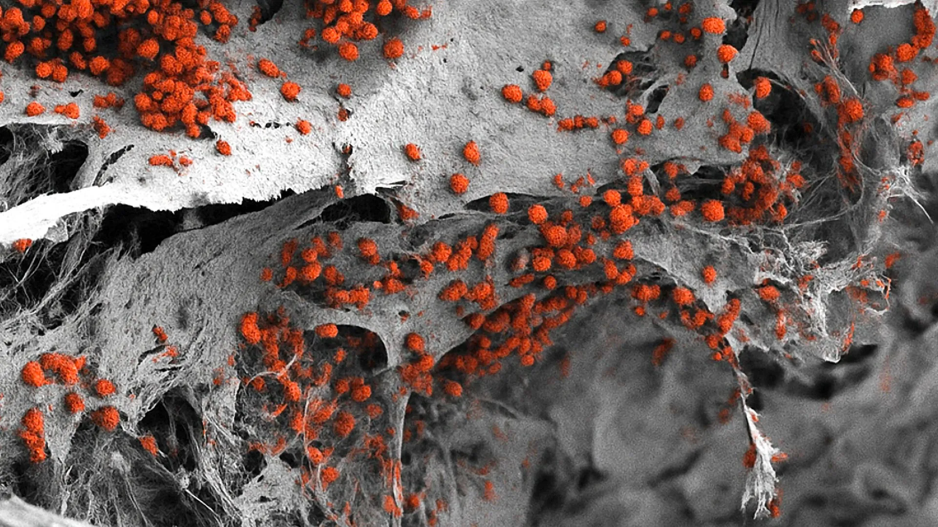
The success of the Basel model is backed by rigorous data confirming its biological activity. Analysis of the three-dimensional structure showed that the laboratory-grown tissue maintained a high degree of cellular diversity and spatial organization similar to that found in biopsies of human bone marrow.
Crucially, the model demonstrated functional hematopoiesis. When human hematopoietic stem cells were introduced into the system, they successfully survived and continued to produce new blood cells for several weeks. This longevity is vital for long-term studies of disease progression or drug toxicity, which often require extended observation periods that simplified models cannot support. The researchers also confirmed the presence of functional blood vessel precursors and nerve-like signals within the niche, marking the first time all these components have been integrated into a single human-derived laboratory system.
Implications for Oncology and Drug Development
The immediate application of this model lies in the field of oncology. Blood cancers are notoriously difficult to treat because malignant cells often "hide" within the protective environment of the bone marrow niches, where they can remain dormant and resistant to chemotherapy. Once treatment ceases, these cells can re-emerge, leading to relapse.
By using the new 3D model, researchers can observe how cancer cells interact with the bone surface and the surrounding blood vessels. This allows for the testing of "niche-disrupting" therapies—drugs designed not just to kill the cancer cells directly, but to strip them of their protective environment, making them more vulnerable to standard treatments.
Furthermore, the pharmaceutical industry stands to benefit significantly. The model offers a more accurate platform for testing drug toxicity and efficacy before human trials begin. As noted by Dr. Andrés García-García, the current size of the model (8mm x 4mm) is ideal for studying complex interactions, though further miniaturization may be required for high-throughput screening, where hundreds of drug compounds are tested simultaneously.
Aligning with the "3Rs" and Ethical Research Standards
Beyond its scientific utility, the development of the human bone marrow model aligns with a global movement in the scientific community known as the "3Rs": Replacement, Reduction, and Refinement of animal experiments. In many regions, including the European Union, there is increasing regulatory and ethical pressure to find alternatives to animal testing.
"Our model brings us closer to the biology of the human organism," stated Professor Ivan Martin. While animal studies have been foundational, they are an approximation. By providing a platform that uses only human cells, the Basel team is contributing to a future where medical research is both more ethically sound and more scientifically accurate. This development is particularly timely given the 2022 passage of the FDA Modernization Act 2.0 in the United States, which allows for the use of alternatives to animal testing, such as cell-based assays and computer models, for drug clearance.
The Future of Personalized Medicine
Perhaps the most ambitious potential for this technology is in the realm of personalized medicine. In the future, a patient diagnosed with a complex blood disorder could have their own cells harvested and used to create a "patient-specific" bone marrow model in the lab. Doctors could then test various chemotherapy cocktails or experimental drugs on that individual’s unique marrow environment to see which treatment is most effective before the patient ever receives a dose.
This approach would eliminate the "trial and error" often associated with cancer treatment, reducing unnecessary side effects and improving survival rates. While the researchers acknowledge that several technical hurdles remain—such as the need to integrate a flowing circulatory system into the model and further reduce the cost of production—the study published in Cell Stem Cell represents the foundational step toward this "biological twin" approach to healthcare.
Conclusion and Scientific Reaction
The scientific community has reacted with cautious optimism to the Basel study. Hematologists have noted that while previous "organ-on-a-chip" technologies offered some insights, they often lacked the "bone" component of the marrow, which is essential for understanding the mechanical stresses and mineral interactions that govern blood production. By including a hydroxyapatite scaffold and iPSC-derived cells, the Basel team has provided the most complete "in vitro" representation of the human bone marrow to date.
As research continues, the team aims to further refine the model by introducing immune system components and exploring how aging affects the marrow niche. The success of this 3D human bone marrow model not only challenges the status quo of animal-centric research but also provides a powerful new lens through which we can view, and ultimately treat, the most complex diseases of the blood.